


Learn the causes of leg swelling and when to worry. Understand key symptoms and seek timely medical advice to manage and treat this common health issue effectively.
Leg Swelling: When to Worry?
Leg swelling is a common symptom that can arise from a variety of causes, ranging from benign to potentially serious health conditions. Knowing when to seek medical advice is crucial in managing this condition effectively. This article will guide you through the important signs that indicate when leg swelling is a cause for concern.
Common Causes of Swelling
Venous insufficiency and Varicose veins
- Poor functioning of the veins can cause blood to pool in the legs, leading to swelling.

Heart failure
- When the heart cannot pump blood effectively, fluid can accumulate in the legs and lungs.
Kidney failure
- Impaired kidney function can lead to fluid retention and swelling in the legs.
Liver Disease
- Liver conditions such as hepatitis and cirrhosis can cause fluid buildup in the legs.
Pregnancy
- As the uterus grows, it puts pressure on the veins in the pelvis and legs, particularly the inferior vena cava, which is the large vein that returns blood from the lower body to the heart. This pressure can slow down blood circulation and lead to fluid buildup in the legs.

Deep Vein Thrombosis (DVT)
- A blood clot in a deep vein can lead to swelling, pain, and redness in the affected leg.

Lymphedema
- Blockage or damage to the lymphatic system can result in fluid retention and swelling.

Medications
- Certain medications, including some blood pressure medications, steroids, and anti-inflammatory drugs, can cause swelling.
Warning Signs of Leg Swelling
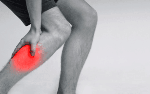

Rapid Onset of Swelling
If you notice that your legs swell up suddenly and severely, it could be a sign of deep vein thrombosis (DVT), especially if the swelling is confined to one leg. In DVT, the clot obstructs blood flow, increasing pressure in the vein. This increased pressure can cause fluid to leak into the surrounding tissues, contributing to swelling and redness. DVT requires immediate medical attention to prevent complications, such as pulmonary embolism.
Pain and Discomfort
Swelling that comes with persistent pain, discomfort, or a feeling of heaviness in the legs should not be overlooked. Pain might indicate underlying issues such as venous insufficiency or a clot due to increased pressure in the veins.
Accompanied by Redness or Warmth
If the swollen area is red and feels warm to the touch, this could be a sign of an infection (cellulitis) or inflammation. Cellulitis is a bacterial infection of the skin and tissues beneath it that can cause inflammation and fluid buildup in the affected area. In DVT, a blood clot formed in the veins can lead to an inflammatory response. Again, obstruction of veins from the clots will also lead to congestion of blood in the leg, contributing to swelling and redness.
Prolonged Swelling Without Improvement
Swelling that does not improve after a few days of home care measures such as elevation, rest, and proper hydration might indicate a more serious condition requiring professional medical evaluation.
Shortness of Breath or Chest Pain
If you experience shortness of breath along with swollen legs, it is crucial to seek immediate medical attention. Leg swelling and shortness of breath are common symptoms of congestive heart failure. The combination of leg swelling and shortness of breath could also be a sign of a pulmonary embolism, which is potentially life-threatening.

When the heart fails to function properly, blood accumulates in peripheral areas, such as the hands and lower legs. This results in the lungs filling with fluid, leading to pulmonary oedema and breathing difficulties.
Fever
If you have a fever along with leg swelling, it could be a sign of a leg infection that has spread throughout your body.
History of Cancer
Cancer cells may spread to the lymph nodes in the groins and pelvis. The nodes themselves can become enlarged, obstructing the lymph vessels, impeding the flow of lymph fluid, and causing swelling (lymphedema). If you have a previous history of cancer, it is important to seek medical attention if you experience leg swelling, as it could be a sign of a recurrence or progression of the disease.
Impact on Daily Activities
When swelling starts to impact your ability to perform daily activities or causes significant discomfort and aches during routine tasks, it is time to consult a healthcare provider.
No Obvious Cause
If you have not been standing or sitting for prolonged periods and there is no clear reason for the swelling, it’s wise to get checked by a doctor. Seeking medical advice in these situations can help diagnose the cause of swelling accurately and allow for appropriate treatment to prevent complications.
Conclusion
Leg swelling is a common issue that affects many people, but understanding the potential causes and knowing when to seek medical advice is key to managing and treating the condition effectively. While mild and infrequent leg swelling can often be managed with lifestyle modifications and simple home remedies, persistent or severe symptoms warrant professional evaluation to rule out more serious conditions. By being proactive about your health and making informed decisions, you can take control of your well-being and keep your legs healthy and functional.

Learn about Deep Vein Thrombosis in the leg: understand its causes, recognise symptoms, and explore prevention techniques to safeguard your vascular health and minimise risks.
Introduction
Deep vein thrombosis (DVT) is a condition that often goes unnoticed, but its potential dangers should not be underestimated. To safeguard your health, it is essential to understand the causes, symptoms, and prevention strategies for DVT in the leg. In this comprehensive guide, we will reveal the mysteries of DVT, shedding light on this debilitating condition.
DVT occurs when blood clots form in deep veins, typically in the legs. If left untreated, these clots can break loose and travel to the lungs, causing a life-threatening condition known as pulmonary embolism (PE). We aim to provide you with the knowledge to identify and address the risk factors associated with DVT, empowering you to take proactive steps towards prevention.
Understanding the Causes of Deep Vein Thrombosis
Deep vein thrombosis (DVT) is a medical condition that can occur due to a combination of genetic and lifestyle factors. Some individuals have a genetic predisposition to develop blood clots, which increases their risk of DVT. This genetic tendency can be passed down from one generation to another, making it crucial to understand your family history to help you identify if you are at higher risk of DVT.
In addition to genetic factors, certain lifestyle choices can contribute to the development of DVT (deep vein thrombosis). Sedentary behaviour, such as sitting or lying down for long periods, can impair blood flow and increase the risk of clot formation. Obesity, smoking, and hormone-based medications like birth control pills can also increase the likelihood of developing DVT.
It is essential to note that while these factors can increase the risk, anyone can develop DVT regardless of their genetic predisposition or lifestyle choices. Understanding the causes can help individuals make informed decisions to mitigate their risk.
Recognizing the Symptoms of Deep Vein Thrombosis
One of the challenges with DVT is that it often presents with little to no symptoms. However, when symptoms do occur, they should not be ignored. The most common symptom of DVT is pain and swelling in the affected leg. The pain may feel like a cramp or a sore muscle, and the swelling may be accompanied by warmth and redness.
Other signs of DVT include a heavy or achy feeling in the leg, skin discolouration, and visible veins. It is important to pay attention to these symptoms, especially if they are persistent or worsen over time. If you experience any of these symptoms, it is crucial to seek medical attention promptly to prevent potential complications.
| Symptoms of Leg DVT | |
| 1 | Swelling of the leg |
| 2 | Pain in the leg, which tends to be worse when standing or walking |
| 3 | Increased warmth in the leg that’s swollen or painful |
| 4 | Red or discoloured skin on the leg |
Complications associated with Deep Vein Thrombosis
Deep vein thrombosis (DVT) can be a severe condition that may result in life-threatening complications. The most significant complication of DVT is pulmonary embolism (PE), which occurs when a blood clot dislodges from the deep veins and moves to the lungs, causing a blockage of blood flow. Symptoms of PE include chest pain, shortness of breath, and, in severe cases, death.
| Symptoms of Pulmonary Embolism | |
| 1 | Unexplained shortness of breath. |
| 2 | Chest pain with deep breathing. |
| 3 | Coughing up blood. |
| 4 | Rapid breathing |
| 5 | Feeling faint |
Apart from PE, DVT can damage the valves in the veins, leading to poor blood flow back to the heart. This condition is known as chronic venous insufficiency (CVI) and can lead to long-term complications called post-thrombotic syndrome (PTS), The symptoms and complications of PTS are similar to those of varicose veins. PTS may affect up to 50% of patients with deep venous thrombosis (DVT) and can have a substantial effect on quality of life. PTS can lead to chronic leg pain, swelling, and skin ulcers due to damage to the veins. It is crucial to detect and treat DVT early to minimise the risks involved and prevent such complications.
| Post Thrombotic Syndrome | |
| 1 | Chronic leg pain, worse with activities e.g. standing and walking |
| 2 | Leg swelling |
| 3 | Leg heaviness or fatigue |
| 4 | Leg cramps |
| 5 | Skin hyperpigmentation/discolouration |
| 6 | Skin thickening |
| 7 | Skin ulceration |
Diagnosing Deep Vein Thrombosis: Tests and Procedures
When DVT is suspected, healthcare professionals employ various tests and procedures to make an accurate diagnosis. One commonly used method is an ultrasound scan, which allows visualisation of the blood flow and clot formation in the deep veins. This non-invasive procedure is painless and provides valuable information for diagnosis.
In some cases, additional imaging tests, such as CT venography or magnetic resonance Venography (MRV), may be performed to provide a more detailed view of the affected veins. These diagnostic tools help healthcare providers determine the appropriate course of treatment.
| Diagnostic Tools | |
| 1 | Ultrasound scan |
| 2 | CT venography |
| 3 | MR venography |
Preventing Deep Vein Thrombosis: Lifestyle Changes and Precautions
Prevention is vital when it comes to DVT, especially for individuals with a higher risk. Making certain lifestyle changes and taking precautions can significantly reduce the likelihood of developing DVT. Simple measures like regular exercise can improve blood flow and reduce clot formation.
For individuals who are at higher risk, such as those with a family history of DVT or those who have undergone surgery, preventive measures may include the use of compression stockings or medication. Compression stockings help improve blood flow in the legs, while medications such as anticoagulants can prevent clot formation.
Additionally, individuals should avoid prolonged periods of sitting or lying down, especially during long flights or car rides. Regular movement and stretching help keep blood flowing properly. Staying hydrated and maintaining a healthy weight are also important factors in reducing the risk of DVT.
For individuals over 60 or those with obesity, incorporating regular physical activity into their daily routine can enhance blood flow and reduce the risk of clot formation. Pregnancy, birth control pills, and hormone replacement therapy (HRT) all influence hormonal balance and clotting risk; women in these categories should discuss their risks and possible preventive strategies with healthcare providers. This might include selecting lower-risk birth control methods and compression stockings during pregnancy and postpartum. For smokers, quitting smoking is a critical step not only for DVT prevention but also for overall health improvement. Lastly, cancer patients should consult with their healthcare team about specific measures to mitigate their risk of DVT, which may include anticoagulant medications or compression stockings.
| Risk Factors | |
| 1 | Family history of DVT |
| 2 | Surgery |
| 3 | Immobility |
| 4 | Age – Older than 60 |
| 5 | Pregnancy |
| 6 | Birth control pills/ Hormone replacement therapy (HRT) |
| 7 | Obese |
| 8 | Smoking |
| 9 | Cancer |
Treatment Options for Deep Vein Thrombosis
The primary goal of treating Deep Vein Thrombosis (DVT) is threefold: firstly, to prevent the clot from growing larger; secondly, to prevent the clot from breaking loose and causing a potentially life-threatening pulmonary embolism (PE); and thirdly, to reduce the chances of future clot formation. Treatment aims to manage immediate risks while minimising long-term complications such as post-thrombotic syndrome, which can lead to swelling, pain, and discolouration in the affected limb. Anticoagulant medication, which thins the blood to prevent clots from enlarging and new ones from forming, is a cornerstone of DVT treatment. In some cases, more invasive procedures may be required, such as:
1) Catheter-directed thrombolysis involves using a catheter to deliver clot-dissolving medication directly to the clot.
2) Thrombectomy, a surgical procedure where the clot is physically removed through a small incision.
3) Insertion of a filter in the inferior vena cava to prevent dislodged clots from getting into the lungs.
Overall, effective DVT management requires a tailored approach that considers the individual’s risk factors, current health status, and potential for adverse effects.
Explore effective strategies for treating ingrown toenails in people with diabetes, ensuring foot health, and preventing complications. Learn essential care tips.
Ingrown Toenails in People with Diabetes
Introduction
Ingrown toenails, also known medically as Onychocryptosis, are a common nail disorder associated with diabetes. Due to their heightened risk of infection and slower healing rates, ingrown toenails can be particularly troublesome for individuals with diabetes. This article will provide an overview of the causes, symptoms, and management strategies for ingrown toenails in people with diabetes.
Causes of Ingrown Toenails
Ingrown toenails occur when the edge of a toenail grows into the surrounding skin. This can lead to pain, redness, and swelling. The big toe is the most likely affected by an ingrown toenail.
Understanding the causes of ingrown toenails is crucial for individuals with diabetes as it can help them take preventive measures. Here are some common causes of this condition:
Improper Trimming: Cutting toenails too short or not straight across can encourage the corners of the nail to dig into the skin.
Tight Shoes: Too tight or narrow footwear can compress the toes, pushing the skin into the nail plate.
Injuries: Any trauma to the toenail, such as stubbing your toe or dropping something heavy on your foot, can cause an ingrown toenail. Activities involving repeated pressure on the toes can also lead to this condition.
Genetics: Some people are born with toenails that are more curved or thick, which makes them more susceptible to becoming ingrown.
Poor Foot Hygiene: Not keeping your feet clean and dry can lead to various foot problems, including ingrown toenails.
Fungal Infections can cause the toenail to become thick or warped, increasing the risk of ingrowth.
The Risks of an Ingrown Nail When You Have Diabetes
In people with diabetes, several factors contribute to the likelihood of developing infection and gangrene of the affected toes:
Poor Circulation (Peripheral Arterial Disease): Diabetes can impair blood flow to the feet, slowing healing and increasing susceptibility to infections.
Nerve Damage (Diabetic Neuropathy): Diabetic neuropathy can significantly reduce foot sensation, which may prevent patients from feeling pain associated with an ingrown toenail. Without the normal pain response, an ingrown toenail may go unnoticed and untreated for a more extended period, increasing the risk of severe infections and complications such as ulcers or even gangrene.
Foot Deformities: Diabetes can lead to changes in foot shape, which might cause abnormal pressure on the toes and nails.

Symptoms to Watch for
Individuals with diabetes must monitor their feet regularly for signs of ingrown toenails, which include:
- Redness and swelling around the nail – The affected area may appear red and swollen due to inflammation caused by the body’s immune response to the infection.
- Pain or tenderness when touching the toe– Diabetic foot wounds can be painful or sensitive. In some cases, patients may experience a loss of sensation in the affected area due to diabetic neuropathy.
- Pus or fluid discharge– Pus or fluid may ooze from the wound, indicating an active infection that requires immediate medical attention.
- Foul smelling– An unpleasant smell from the wound may indicate a severe infection.
Grading of the Severity of Ingrown Toenails
Understanding the grading of the severity of ingrown toenails is crucial for effective treatment. Ingrown nails typically progress through three stages:
Mild (Stage 1):
Redness, swelling, and pain are usually present along one or both sides of the nail (lateral nail fold).

Moderate (Stage 2):
As the condition worsens, the symptoms become more pronounced. The affected area may become more swollen and painful with discharge, indicating a bacterial infection.

Severe (Stage 3):
In the most advanced stage, the ongoing infection and worsening symptoms can lead to the overgrowth of granulation tissue around the toenail.
An abscess may develop when the infection spreads to a broader area of the affected toe, requiring more intensive treatment, possibly including surgery.

Preventive Measures
Preventing ingrown toenails is particularly important for people with diabetes. Some preventive measures include:
Proper Nail Trimming:
Cut toenails straight across without trimming the corners, and avoid cutting them too short to prevent ingrown nails.


Appropriate Footwear:
Shoes that fit well and provide enough room around the toes can help prevent pressure on the nails.
Regular Foot Checks:
These are vital parts of self-care for individuals with diabetes. A daily inspection of the feet can help catch early signs of problems, including ingrown toenails, before they escalate.
Conservative Management
Conservative management is highly effective for early-stage ingrown toenails.
Elevation of the ingrowing nail edge:
Use a pair of forceps to pack a small piece of cotton wool under the free corner of the nail. Similarly, one can also use dental floss or a gutter splint to place under the edge of the nail to prevent it from digging into the surrounding tissue.

Elevation of ingrown nail edge with cotton

Elevation of ingrown nail edge with gutter splint

Elevation of ingrown nail edge with dental floss
Topical Antibiotics/antiseptics:
Applying antibiotic / antiseptic ointment to the affected area can help prevent infection and promote healing.
Pain Relief:
Use over-the-counter painkillers, such as Nonsteroidal anti-inflammatory drugs (NSAIDs) and paracetamol, to help reduce pain and inflammation.
When to See a Doctor
Individuals with diabetes should take ingrown toenails seriously, as even minor foot injuries can lead to severe complications. These complications include the risk of developing gangrene, which could result in the loss of a limb. Poor circulation, common in diabetes, further increases the likelihood of infection and impedes healing, making immediate medical attention crucial.
If you develop a foot wound because of an ingrown toenail, you should never attempt self-treatment. Early specialist intervention can prevent serious complications.
Managing the symptoms and seeking a permanent solution to prevent recurrence is crucial.
Professional Management and Treatment
Assessment for circulation:
Assessment of blood supply to the foot in people with diabetes before undergoing any toe surgery or treatment is crucial because of the risk of compromised circulation often associated with diabetes. Poor blood supply can significantly increase the risk of infection and gangrene following surgical interventions.
Surgical options:
There is still no agreement on the most effective treatment for ingrown toenails. Managing ingrown toenails can be tricky, and the best treatment often depends on the severity and frequency of the problem. Here are some common approaches:
Simple nail avulsion/wedge excision (Not recommended)
Both nail avulsion and wedge excision are common procedures used to treat ingrown toenails. During these procedures, the part of the nail causing the ingrown toenail is temporarily removed. However, the nail matrix, the tissue under the skin that produces the nail, is not treated. If the nail matrix is not treated, the nail will likely grow back in the same pattern, potentially leading to the same ingrown condition. Therefore, it is essential to treat the nail matrix to prevent the recurrence of an ingrown toenail.
Partial Nail Avulsion with Phenol Matricectomy
This is one of the most common procedures. It involves removing a portion of the toenail (of the affected side) and applying a chemical called phenol to the nail matrix. The phenol destroys the matrix cells, preventing regrowth of the nail at the treated site and thereby reducing the chance of recurrence. This treatment is highly effective, with a high success rate and low recurrence of ingrown toenails.
Wedge Resection with Surgical Matricectomy (Zadik’s Procedure)
This surgical technique removes a wedge-shaped portion of the nail and the underlying nail bed and matrix (nail root). This method is highly effective for preventing recurrence and is often used when the ingrown toenail is recurrent or severe.
Complete Nail Avulsion with Matricectomy:
In more severe cases, the entire nail may be removed, followed by matricectomy. This is a less common procedure for ingrown toenails unless the problem is extensive. Complete nail avulsion can be followed by matricectomy if there is a high risk of recurrence.
Debridement and drainage of pus:
In cases associated with severe infection, debridement and drainage of pus may need to be carried out before proceeding to any definitive nail procedure. Antibiotics are often required in this situation.
Conclusion
Effective management of ingrown toenails is crucial for individuals with diabetes as part of their overall foot care. Regular foot inspections, preventive measures, and prompt treatment are key to avoiding complications and maintaining good foot health. Those with diabetes should work closely with their healthcare providers to manage their foot health and prevent problems such as ingrown toenails.
Diabetic neuropathy increases the risk of foot ulcers by reducing sensation, making it hard to detect injuries that can lead to gangrene and serious infections.
Introduction
Diabetic neuropathy is a common and serious complication of diabetes, affecting up to 50% of individuals with diabetes during their lifetime. It encompasses a group of nerve disorders caused by diabetes, which can significantly impair quality of life and increase the risk of further complications, such as diabetic foot ulcers. This article explores the types of diabetic neuropathy—sensory, motor, and autonomic—and the development of diabetic foot ulcers, providing essential insights for management and prevention.
What is Diabetic Neuropathy?
Diabetic neuropathy refers to the damage inflicted on nerves due to high blood sugar levels associated with diabetes. Over time, elevated glucose levels can deteriorate the small blood vessels that supply vital nutrients and oxygen to the nerves, leading to neuropathy. This condition can manifest in various forms, primarily categorized into sensory, motor, and autonomic neuropathy, each affecting different nerve groups and leading to distinct complications.
1. Sensory Neuropathy
Sensory neuropathy affects the nerves responsible for sensation. It is the most common form of neuropathy in people with diabetes. It typically starts in the feet and can spread to the hands. Symptoms include numbness, tingling, pain, or weakness in the affected areas. The loss of sensation means that injuries, cuts, or blisters can go unnoticed, setting the stage for severe infections or ulcers.

Sensory Neuropathy with Lack of Pain Sensation
2. Motor Neuropathy
Motor neuropathy involves the nerves that control muscle movement. When these nerves are damaged, muscles might not function properly, leading to muscle weakness and imbalances in the foot’s structure. This can affect a person’s balance and coordination, increasing the risk of falls and injuries. Furthermore, muscle weakness can contribute to deformities in the feet, such as claw toes, hammertoes, or the collapse of the foot arch, which alter foot biomechanics and, thus, abnormal gait. These deformities can also change the way weight is distributed across the foot, leading to abnormal pressure points when standing, walking, or running. These abnormal pressure points of the foot are typically the ball of the foot or the underside of the toes to endure excessive stress. Over time, this stress over the pressure areas can lead to callus formation and, eventually, skin breakdown beneath these hardened areas, forming ulcers.

Hammer Toe Deformity

Claw Toe Deformity

Calluses on Pressure Points

Claw Toes: Pressure Points and Ulcers

Flat Foot: Pressure Point

High-arch Foot: Pressure Points
3. Autonomic Neuropathy
Autonomic neuropathy affects the nerves that control involuntary bodily functions, including heart rate, digestion, and bladder control. Relevant to foot health, autonomic neuropathy can impair sweat and oil production in the feet. This results in dry, cracked skin, which is more susceptible to infection and the development of ulcers.

Diabetic Autonomic Neuropathy: Dry Skin

Diabetic Autonomic Neuropathy: Dry and Cracked Skin
Diabetic Foot Ulcers: A Dangerous Complication
Diabetic foot ulcers are open sores or wounds that typically occur on the bottom of the foot. In individuals with diabetes, these ulcers often result from a combination of neuropathy, poor circulation (another complication of diabetes), and an inability to heal properly. Foot deformities and poor footwear can also create constant pressure points and contribute to the formation of ulcers.

Diabetic Foot Ulcers
Reduced Blood Flow (Peripheral Arterial Disease)
Although not a direct result of neuropathy, many individuals with diabetes also suffer from peripheral arterial disease (PAD), which reduces blood flow to the feet. Reduced blood flow impairs healing and increases the risk of ulcers becoming infected, compounding the problem initiated by neuropathy.
Here are some hard facts concerning diabetic PAD:
• 70% of diabetic wounds/ ulcers involve PAD.
• Almost 85% of amputations related to diabetes are preceded by ulcers, indicating that many of these amputations could be prevented.
• Two-thirds of older adults who undergo amputation fail to regain their independence.
• Approximately 70% of individuals who undergo amputation pass away within five years.

Diabetic Foot Gangrene

Diabetic Foot Gangrene with Infection
Risk Factors and Prevention
Key risk factors for diabetic foot ulcers include:
1. Duration of Diabetes: Longer duration increases the risk.
2. Poor Blood Glucose Control: High blood sugar levels can lead to nerve damage and poor circulation.
3. Diabetic Neuropathy: Nerve damage that causes loss of sensation in the feet.
4. Peripheral Arterial Disease (PAD): Reduced blood flow to the feet, impairing wound healing.
5. Foot Deformities: Such as hammertoes, bunions, or Charcot joints, which can create pressure points.
6. Inappropriate Footwear: Shoes that do not fit well can cause blisters, sores, and ulcers.
7. Previous Foot Ulcers: History of foot ulcers increases the likelihood of future occurrences.
8. Poor Foot Hygiene and Care: Neglecting foot care can lead to conditions that promote ulcer formation.
9. Smoking: Impairs circulation, exacerbating risks associated with poor blood flow.
10. Obesity: Increases pressure on the feet and complicates diabetes management.
Preventive measures
Preventive measures for diabetic foot ulcers are crucial for maintaining foot health and avoiding complications. Here are key strategies to consider:
1. Regular Foot Inspections: Check your feet daily for cuts, blisters, redness, swelling, or nail problems.
2. Manage Blood Glucose Levels: Maintain your blood sugar within the recommended range to help prevent further nerve damage and improve healing.
3. Wear Appropriate footwear: Choose well-fitting, comfortable shoes that provide good support and cushioning. Avoid tight shoes and pointed toes.
4. Keep Feet Clean and Moisturized: Wash your feet daily and dry them gently, especially between the toes. Apply a moisturizer to prevent dry skin from cracking.
5. Trim Nails Carefully: Cut nails straight across and file the edges. Avoid cutting nails too short to prevent injury.
6. Avoid Walking Barefoot: Always wear shoes or slippers, even indoors, to avoid injuries.
7. Stop Smoking: Smoking impairs circulation and reduces the amount of oxygen that reaches the feet, which can hinder wound healing.
8. Regular Check-ups with Healthcare Provider: Have your feet examined by a healthcare professional at least once a year – more often if you have known risk factors for foot problems.
9. Exercise Regularly: Physical activity can improve circulation and help maintain healthy blood sugar levels.
10. Educate Yourself and Family: Learn about the risks and preventative care for diabetic foot issues. Educate family members so they can help with inspections and recognize early signs of complications.
Implementing these measures can significantly reduce the risk of developing foot ulcers and ensure early detection and treatment of potential problems.
Conclusion
Diabetic neuropathy and the resulting risk of foot ulcers are significant concerns for individuals with diabetes. Understanding the different types of neuropathy and their implications can help in early identification and appropriate management. By integrating comprehensive foot care into the daily routine and maintaining optimal control of blood glucose levels, individuals with diabetes can significantly reduce their risk of developing serious foot complications. Remember, early intervention and consistent management are key to preventing the severe consequences of diabetic neuropathy.
Discover the benefits of sclerotherapy for spider vein treatment: a minimally invasive, effective solution with minimal discomfort and high success rates.
Introduction
Say goodbye to those unsightly spider veins with an effective treatment solution: Sclerotherapy. If you’re tired of hiding your legs or feeling self-conscious about those visible veins, you’re not alone. Many are in search of effective ways to address these veins, and fortunately, a reliable option exists that could help restore your skin’s smoother, more uniform appearance.
Sclerotherapy presents itself as a minimally invasive approach, offering a method to reduce the visibility of spider veins. Sclerotherapy for spider veins is also termed microsclerotherapy, as the procedure involves using a very fine needle on very small veins. The procedure involves carefully injecting a specialized solution into the targeted veins, which encourages them to collapse and be gradually absorbed by the body, resulting in clearer skin. Patients typically find spider vein treatment minimally uncomfortable yet effective in reducing visibility.
Sclerotherapy improves the aesthetic appearance of your legs and provides relief from associated symptoms such as itching, burning, and discomfort. This non-surgical solution provides a straightforward path to reducing spider vein visibility without extensive downtime.
If spider veins have been a source of concern for you, considering sclerotherapy could be a step towards feeling more comfortable and confident in your skin. Scheduling a consultation could be your initial move towards understanding how sclerotherapy might fit into your journey towards smoother, more vein-free legs.
Understanding the Causes and Symptoms of Spider Veins
Spider veins, also known as telangiectasias, are small, dilated blood vessels that appear close to the surface of the skin. They often resemble spider webs or branches of a tree, hence the name. While they can occur anywhere on the body, they are most commonly found on the legs and face. Spider veins are usually harmless, but they can cause discomfort and self-consciousness.

Several factors contribute to the development of spider veins, including genetics, hormonal changes, pregnancy, obesity, prolonged standing or sitting, and age. Hormonal fluctuations, such as those that occur during pregnancy or menopause, can weaken the vein walls and lead to their dilation. Additionally, as we get older, the valves in our larger veins might not work as well as they used to. These valves help keep blood flowing in the right direction. When they weaken, pressure can build up in the veins. This increased pressure can then spread to smaller veins, including those close to the skin’s surface, causing spider veins to form.
The symptoms of spider veins can vary from person to person. Many individuals experience no symptoms other than the visible appearance of the veins. However, others may experience itching, burning, or aching sensations in the affected area. In some cases, spider veins may cause discomfort, especially after prolonged periods of standing or sitting. If you’re experiencing any of these symptoms or are simply unhappy with the appearance of spider veins, sclerotherapy may be the solution you’ve been seeking.
How Sclerotherapy Works to Treat Spider Veins
Sclerotherapy is a highly effective treatment for spider veins. It works by injecting a special solution, known as a sclerosant, directly into the affected veins. This solution irritates the vein walls, causing them to collapse and eventually fade away. Over time, the body absorbs the treated veins, resulting in smoother, clearer skin. The most commonly used sclerosants for this procedure include sodium tetradecyl sulphate (STS), polidocanol (also known as laureth-9 or Asclera), and hypertonic saline solution. They are all approved by the FDA for the sclerotherapy treatment of spider veins.


During the sclerotherapy procedure, a fine needle will be used to carefully inject the sclerosant into the spider veins. The number of injections required will depend on the size and extent of the veins being treated. The procedure itself is relatively painless, with most patients experiencing only minimal discomfort. Some individuals may feel a slight burning or stinging sensation at the injection site, but this typically subsides quickly.
After the sclerosant is injected, compression stockings or bandages will be used to apply pressure to the treated area. This helps to keep the collapsed veins closed and aids in the absorption process. The length of the sclerotherapy session can vary depending on the number of veins being treated, but most procedures are completed within 30 minutes to an hour.

When it comes to treating spider veins, there are several options available. However, sclerotherapy stands out as the best treatment solution for many reasons.
One of the main advantages of sclerotherapy is its effectiveness. Studies have shown that sclerotherapy can achieve a success rate of up to 80-90% in reducing the appearance of spider veins. Unlike some other treatments, such as laser therapy, which may require multiple sessions for optimal results, sclerotherapy often produces noticeable improvements after just one treatment.
Another benefit of sclerotherapy is its versatility. Many people who have spider veins also have concomitant reticular veins, which are a bit larger. The procedure can be performed on both spider veins and larger veins, making it suitable for a wide range of cases.
Sclerotherapy is also a minimally invasive procedure, meaning it does not require surgery. This reduces the risks associated with more invasive treatments and eliminates the need for general anaesthesia. Most patients can resume their normal activities immediately after the procedure, although strenuous exercise and hot baths or saunas should be avoided for a short period of time.
Furthermore, sclerotherapy provides relief from associated symptoms, such as itching, burning, and discomfort. By collapsing the affected veins, the procedure alleviates the pressure and congestion that can cause these symptoms. This not only improves the aesthetic appearance of the legs but also enhances overall comfort and well-being.
Benefits of Choosing Sclerotherapy over Other Treatment Options
When it comes to treating spider veins, there are several options available. However, sclerotherapy stands out as the best treatment solution for many reasons.
One of the main advantages of sclerotherapy is its effectiveness. Studies have shown that sclerotherapy can achieve a success rate of up to 80-90% in reducing the appearance of spider veins. Unlike some other treatments, such as laser therapy, which may require multiple sessions for optimal results, sclerotherapy often produces noticeable improvements after just one treatment.
Another benefit of sclerotherapy is its versatility. Many people who have spider veins also have concomitant reticular veins, which are a bit larger. The procedure can be performed on both spider veins and larger veins, making it suitable for a wide range of cases.
Sclerotherapy is also a minimally invasive procedure, meaning it does not require surgery. This reduces the risks associated with more invasive treatments and eliminates the need for general anaesthesia. Most patients can resume their normal activities immediately after the procedure, although strenuous exercise and hot baths or saunas should be avoided for a short period of time.
Furthermore, sclerotherapy provides relief from associated symptoms, such as itching, burning, and discomfort. By collapsing the affected veins, the procedure alleviates the pressure and congestion that can cause these symptoms. This not only improves the aesthetic appearance of the legs but also enhances overall comfort and well-being.
The Sclerotherapy Procedure: What to Expect During and After Treatment
The sclerotherapy procedure typically takes place in a clinic. Here’s what you can expect during and after the treatment:
1. Preparation: Before the procedure begins, you will be asked to lie down on a treatment table, and the healthcare professional will cleanse the area being treated. They may mark the veins with a pen to guide them during the injection process.
2. Injection: A fine needle will be used to carefully inject the sclerosant solution into the spider veins. You may feel a slight pinch or stinging sensation as the needle is inserted, but this should subside quickly. Multiple injections may be necessary, depending on the number and size of the veins being treated.
3. Compression: After the injections are complete, compression stockings or bandages will be applied to the treated area. This helps prevent blood from flowing back into the collapsed veins and encourages healing. You will be instructed to wear these compression garments for a specified period of time following the procedure.
4. Recovery: Following the procedure, you will be able to resume your normal activities immediately. However, it’s important to avoid strenuous exercise, hot baths or saunas, and direct sun exposure for a period of time. Some individuals may experience mild bruising, swelling, or redness at the injection sites, but these symptoms usually resolve on their own within a few days to a few weeks.
5. Results: Over time, you will begin to notice the spider veins fading away. It typically takes a few weeks to several months for the full results to become visible, as the body gradually absorbs the treated veins. In some cases, multiple sclerotherapy sessions may be required to achieve the desired outcome.
By following these guidelines and closely following your healthcare professional’s instructions, you can ensure a smooth and successful recovery from sclerotherapy.
Risks and Side Effects of Sclerotherapy for Spider Veins
Like any medical procedure, sclerotherapy carries certain risks and potential side effects. While complications are relatively rare, it’s important to be aware of them. Here are some potential risks associated with sclerotherapy:
- Allergic reactions: Some individuals may experience an allergic reaction to the sclerosant solution. This can manifest as hives, itching, difficulty breathing, or swelling of the face, lips, or tongue. If you experience any of these symptoms, seek immediate medical attention.
- Skin discolouration or hyperpigmentation: Temporary skin discolouration is a common side effect of sclerotherapy. The treated veins may initially appear darker before fading away. In some cases, hyperpigmentation or brown spots may develop at the injection sites. These discolourations usually resolve on their own within a few months.
- Formation of new vessels or matting: This complication can occur when the body responds to the destruction of leg veins by creating new, smaller veins near the area treated with sclerotherapy. Often, these newly formed veins will fade and vanish on their own without the need for further treatment over a period of a few months.
- Skin ulceration: In rare instances, sclerotherapy may cause skin ulceration. This occurs when the sclerosant solution leaks into the surrounding tissue, leading to tissue damage. Skin ulcerations may require additional treatment and can result in scarring.
- Infection: While infection is rare after sclerotherapy, it can occur. It’s important to keep the treated area clean and follow any post-treatment care instructions provided by your healthcare professional.
- Blood clot formation: Deep Vein Thrombosis (DVT) is very rare for sclerotherapy on spider veins when it is performed by a qualified healthcare professional.
It’s essential to discuss any concerns or questions you may have with your healthcare professional before undergoing sclerotherapy. They will provide you with detailed information about the potential risks and side effects specific to your case.
Frequently Asked Questions About Sclerotherapy for Spider Veins
1. Is sclerotherapy for spider veins painful?
Sclerotherapy is generally well-tolerated and causes minimal discomfort. Most individuals report feeling only a slight pinch or stinging sensation during the injections. Any discomfort experienced during the procedure is usually temporary and subsides quickly.
2. How long does sclerotherapy take to work?
The timeline for seeing results from sclerotherapy varies from person to person. While some individuals may notice improvements within a few weeks, others may require several months to see the full effects. It’s important to be patient and allow your body time to absorb the treated veins fully.
3. How many sclerotherapy sessions will I need?
The number of sclerotherapy sessions required depends on several factors, including the size and number of spider veins being treated and your individual response to the treatment. Some individuals achieve the desired results after a single session, while others may require multiple sessions spaced several weeks or several months apart for optimal outcomes.
4. Does insurance cover sclerotherapy?
In general, sclerotherapy is not covered by insurance if it is carried out for cosmetic reasons. However, it may be covered by insurance if it is deemed medically necessary when spider veins cause significant symptoms like pain, aches, tiredness, and feeling heavy in the legs. However, coverage varies depending on your insurance provider and policy. It’s advisable to check with your insurance company beforehand to determine if sclerotherapy is covered and what documentation or requirements are needed.
5. Can sclerotherapy be performed on varicose veins?
Sclerotherapy is primarily used to treat spider veins. However, it can also be effective in treating small varicose veins. For larger varicose veins, other treatment options, such as endovenous laser therapy or surgical procedures, may be more suitable. It’s best to consult with a healthcare professional to determine the most appropriate treatment for your specific case.
Conclusion: Sclerotherapy is an Effective Solution for Spider Veins
Sclerotherapy offers an effective solution if you are tired of hiding your legs or feeling self-conscious about those visible spider veins. This minimally invasive procedure has a high success rate in reducing spider veins’ appearance and relieving associated symptoms.
Don’t let spider veins hold you back any longer. Take the first step towards smoother, vein-free legs by booking a consultation with Dr Tan Yih Kai, an experienced vascular surgeon with over 20 years of specialized experience in sclerotherapy. Your action could be the transformative step you’ve been searching for.
Iliac Vein Compression Syndrome (IVCS), also known as May-Thurner syndrome, was first identified in 1956 by two doctors, May and Thurner. It happens when a vein on the left side of the body, called the left common iliac vein, gets compressed by an artery from the right side, known as the right common iliac artery. Over time, this constant squeezing can cause the vein to become narrow or even blocked because of scar tissue and other changes. This can make it hard for blood to flow through the vein as it should. This explains why deep vein thrombosis (DVT), blood clots in deep veins, is five times more common in the left versus right leg.


In fact, the iliac vein may also be compressed on the right in some people. The exact number of people who have iliac vein compression isn’t known. This condition can affect both men and women. About 30% of people might have it without showing any symptoms, a situation sometimes called a “permissive lesion.” Essentially, symptoms will start if something else goes wrong, like an infection, or an injury to the leg.

Causes of leg swelling:
Swelling in the legs can happen for many reasons, and it’s important to know why it’s happening to get the right treatment. Here’s a simpler way to understand the causes:
Conditions that will cause bilateral leg swelling only:
- Heart failure
- Kidney failure
- Liver failure
- Side effects from certain medicines.
Condition that will cause either one or both legs swelling:
- Varicose veins, which are swollen, twisted veins.
- Blockage or clot in the deep veins of the legs (known as DVT).
- Iliac vein compression syndrome, where a vein in the pelvis is squeezed.
- Lymphedema, a condition where fluid builds up because the lymphatic system is blocked or damaged.
If your legs are swollen, it’s crucial to see a specialist, like a vascular surgeon, who can determine what’s causing the swelling. Dr. Tan Yih Kai is an experienced Vascular Surgeon who specializes in treating leg swelling caused by vein problems. Don’t hesitate to contact him for advice and to schedule an appointment.
Symptoms:
People with iliac vein compression may develop symptoms because of increased pressure in the veins. The symptoms include:
- Formation of blood clots in the iliofemoral vein, which is a major vein in the leg (known as DVT, or deep vein thrombosis).
- Swelling in the leg
- Pain in the leg.
- Ulcers or open sores that don’t heal easily.
- Lipodermatosclerosis, a type of skin change where the skin becomes thickened and discoloured, often looking like it’s tightening.
- Venous claudication, which means leg pain or discomfort, especially when walking, due to poor blood flow.
These symptoms occur because of upstream vein compression in the pelvis, which leads to higher pressure in the leg veins, making it hard for blood to flow properly.
Diagnostic Evaluation:
For diagnosing iliac vein compression, the following evaluations are commonly used:
Ultrasound: This test checks for varicose veins or venous reflux, a condition in which blood flows backwards in the veins.
CT venogram or MR venogram: These are specialized scans that help visualize compression in the iliac vein and check for the presence of tumours in the pelvic area.
Diagnostic Venogram: This is a type of X-ray used to visualize the veins. During the procedure, a special dye is injected into the veins, making them visible on X-ray images. However, it only provides 2D images and might not catch everything.

Endovascular Interventions
Endovascular interventions, which involve treating from within the blood vessels, have become the preferred method of treatment.
Using a stent to open up the veins in the pelvis and lower abdomen (specifically the iliac and caval veins) has proven to be both safe and effective. This approach is typically recommended for patients who have ongoing, noticeable symptoms from deep vein problems that haven’t improved with standard treatments.
A tool called Intravascular Ultrasound (IVUS) is commonly used during the stenting procedure to help place the stent accurately and ensure the best possible outcome.

IVUS allows the visualization of:
- Percentage of vein stenosis
- Real-time vessel diameter measurement
- Length of stenosis
- Location of side branches
- Calcium, thrombus, and intravenous scarring


What to expect after a stent in the iliac vein?
The stent’s placement should only result in mild discomfort that subsides quickly. It’s common for some patients to feel back pain for about 1 to 2 weeks after the procedure. Typically, this discomfort can be effectively managed with over-the-counter painkillers or anti-inflammatory medications.
The outcome of iliac vein stenting
Research on iliac vein stenting has found very promising results. The success rate of the stent remaining open and effective was found to be 98.7% after 6 months, 98.3% after 1 year, and 97.9% after 2 years of follow-up.
Conclusion:
Chronic venous disease can seriously impact a person’s life. Often, iliac vein compression syndrome isn’t diagnosed as frequently as it should be. Venous stenting, a procedure with a low risk of complications, has a very low chance of causing serious problems. It’s not only a safe and effective treatment, but it also offers lasting results.
FAQs about Iliac Vein Compression Syndrome
What is the primary cause of Iliac Vein Compression Syndrome (IVCS)?
The primary cause of IVCS is the anatomical compression of the left common iliac vein by the right common iliac artery. This pressure can lead to the vein’s narrowing or occlusion, restricting blood flow and increasing the risk of blood clots and swelling in the legs.
Can lifestyle changes improve IVCS symptoms?
Yes, lifestyle changes such as regular exercise, maintaining a healthy weight, and avoiding long periods of sitting or standing can improve blood flow and reduce the symptoms associated with IVCS.
What are the long-term effects of a stent for IVCS?
The placement of a stent can provide immediate relief from IVCS symptoms. Long-term effects typically include improved blood flow and a significant reduction in leg swelling and pain. Stents have a high patency rate, meaning they remain open and effective for extended periods after implantation.
Is there a genetic predisposition to IVCS?
There is no clear genetic predisposition to IVCS.
How does pregnancy affect IVCS?
Pregnancy can exacerbate IVCS due to the increased pressure on the pelvic veins and the higher volume of blood in the body. Pregnant women may experience more significant swelling and discomfort if they have IVCS.
What are the latest advancements in IVCS treatment?
The most significant advancements in IVCS treatment include specially designed iliac vein stents, intravascular ultrasound (IVUS)-guided interventions to allow precise placement of stents, and the development of new, less invasive endovascular techniques.
How does IVCS cause leg swelling?
The compression in IVCS can hinder venous return from the legs to the heart, leading to increased venous pressure, which can cause fluid to accumulate in the legs, resulting in swelling.
Can IVCS be treated without surgery?
Mild cases of IVCS can sometimes be managed with compression stockings, exercise, and medications to improve blood flow. However, severe cases often require endovascular procedures like angioplasty or stenting.
Is IVCS a lifelong condition?
IVCS can be a lifelong condition if not treated. However, with appropriate management, including surgery if necessary, many patients can expect a return to normal activities and minimal symptoms.
If leg swelling persists or if you suspect IVCS, seeking the expertise of a vascular surgeon is imperative. Dr. Tan Yih Kai is an experienced Vascular Surgeon who specializes in treating leg swelling caused by vein problems. Don’t hesitate to contact him for advice and to schedule an appointment.
Discover the latest advancements in varicose veins treatments that are changing the narrative from painful surgeries to minimally invasive, effective solutions. Learn from Dr. Tan at The Vascular Vein and Wound Centre in Singapore about how these evidence-based treatments provide a safer, more comfortable alternative to traditional vein stripping surgery, addressing the root cause of venous insufficiency with state-of-the-art technology.
Varicose Veins and Treatments
Fortunately, the daunting tales of leg vein surgery belong to the past. Today, there are numerous effective and minimally invasive varicose veins treatment methods available that cause minimal discomfort and require a shorter recovery time.
Varicose veins originate from an underlying condition known as venous insufficiency. Both genetic factors and lifestyle choices play a role in its onset. Symptoms of discomfort and pain arise when the small, one-directional valves in the veins are overstressed and start to fail. This failure can cause blood to flow in the reverse direction and pool towards the ankle, leading to the veins enlarging over time and forming varicose veins. This condition is most frequently seen in the lower parts of the body, such as the legs, ankles, and feet.


Contrary to the common misconception that varicose veins are merely a cosmetic concern, they can lead to various problematic symptoms, including swelling in the legs, a sensation of heaviness, muscle cramps, itching, and fatigue. In some cases, the symptoms can be severe enough to adversely affect one’s quality of life. If venous insufficiency, the condition underlying varicose veins, is not addressed, it could predispose individuals to serious health issues such as blood clots and venous ulcers, which are open wounds that do not heal.
If you’re thinking about undergoing a procedure to eliminate varicose veins, it’s important to fully investigate all your treatment possibilities. Consulting with a vein specialist will allow for assessing your symptoms and providing tailored treatment suggestions. Meanwhile, here is essential information you should be aware of regarding varicose vein treatment. At The Vascular Vein and Wound Centre, Dr Tan, a leading vein specialist in Singapore, provides a wide range of proven, effective, and minimally invasive treatments for varicose veins that can quickly alleviate symptoms, resulting in significantly less discomfort and a quicker recovery period.
1. Varicose vein stripping surgery
Nowadays, the practice of varicose vein stripping surgery has become quite uncommon, primarily because the recovery process can be quite discomforting and may last up to four weeks. With the availability of far superior alternatives, undergoing a painful surgery with a lengthy recovery period seems unnecessary now.
2. The modern varicose vein treatment options
All the varicose vein treatments offered in the centre are evidence-based which means the device is safe and effective for its intended use.
a) Endovenous Radiofrequency Ablation Therapy
During Radiofrequency Ablation (RFA) treatment, a vein specialist will insert a thin catheter into the affected vein under ultrasound guidance. Radiofrequency (RF) energy is then used via the RF catheter to heat and seal the problematic vein. Right after the procedure, the blood flow is redirected to nearby healthy veins before flowing back to the heart. The body gradually absorbs the closed vein and scars it over.


b) Mechanochemical ablation (MOCA)
Mechanochemical ablation (MOCA), also referred to as ClariVein is a minimally invasive procedure used to treat varicose veins. It does not use heat energy to seal vein. It combines two techniques: mechanical disruption of the vein wall using a rotating wire or catheter and the chemical effect of a sclerosant solution, which is injected into the vein. This dual-action approach enhances the effectiveness of the treatment.
A catheter is inserted into the affected vein under ultrasound guidance during the MOCA procedure. The catheter delivers a combination of mechanical agitation and a sclerosant solution, causing the vein to collapse and seal shut. Over time, the treated vein is absorbed by the body, and blood flow reroutes to healthier veins.
Because MOCA does not use heat energy like RFA, there is less risk, albeit low, of heat-related damage to surrounding nerves. This treatment method is especially suitable for veins below the knee as nerves may run close to the veins.

Step 1. The ClariVein infusion catheter is inserted into the vein through the skin under ultrasound guidance.

Step 2. The motorised handle of the ClariVein catheter is utilised to rotate the catheter tip while pulling it back. This rotation induces vessel spasm, hindering forward flow and allowing for a concentrated infusion of liquid sclerosant into the targeted area.
c) Ultrasound Guided Foam Sclerotherapy
Ultrasound-Guided Sclerotherapy represents yet another minimally invasive method aimed at sealing off dysfunctional varicose veins and redirecting venous blood circulation. This procedure utilizes ultrasound imaging to identify the problematic vein. Upon identification, a special medication (sclerosant) is administered through injection, which irritates the vein, leading to its closure. Like our other treatments, this method typically requires minimal to no downtime for recuperation.

The advantages of minimally invasive treatment modalities
The advantages of minimally invasive treatment modalities over traditional vein stripping surgery:
1. Reduced Pain: Minimally invasive procedures typically involve smaller incisions or needle punctures, resulting in less trauma to the surrounding tissues and reduced postoperative pain compared to vein stripping surgery.
2. Faster Recovery: Patients undergoing minimally invasive treatments generally experience shorter recovery times and can return to normal activities more quickly than those undergoing vein stripping surgery, which may require more extensive recovery periods.
3. Lower Risk of Complications: Minimally invasive techniques are associated with lower rates of complications such as infection, bleeding, and wound healing issues compared to traditional surgery.
4. Minimal Scarring: Because minimally invasive procedures involve smaller incisions or puncture sites, they often result in minimal scarring, leading to better cosmetic.
Systemic factors
The wound healing process is a complex series of biological events that involve various cellular and molecular interactions. Our bodies aim to restore the damaged tissues and regain their structural and functional integrity through this process. Understanding the myriad of factors that affect wound healing is crucial for effective care and speedy recovery. This article delves into the complexities of wound healing, providing insights and guidance on how various factors play a role in this intricate process.
Several systemic factors can influence the wound healing process. These include:
- Age: As we get older, the healing process tends to slow down due to reduced cellular activity and decreased collagen production.
- Chronic diseases: Chronic diseases, such as diabetes, cardiovascular diseases, and autoimmune disorders, can impair wound healing by affecting blood circulation and immune function.
- Nutrition: Adequate nutrition, including a balanced diet with sufficient protein, vitamins, and minerals, is essential for optimal wound healing.
- Medications: Certain medications, such as immunosuppressants and steroids, can interfere with the healing process.
Local factors
In addition to systemic factors, local factors at the wound site can also impact healing:
- Infection: Wounds that become infected may experience delayed healing or complications. Proper wound care with antibiotics is crucial to prevent further complications.
- Wound size and depth: Large, deep wounds or wounds with significant tissue loss may require more time and interventions to heal properly.
- Mechanical stress: Excessive tension, pressure, or movement at the wound site can impede healing. The location of the wound, such as over joints or bony prominences, can also affect the healing process.
- Poor blood supply: Wounds in areas with compromised blood flow, such as in individuals with peripheral arterial disease, may heal more slowly due to reduced oxygen and nutrient delivery.
Gain insights into the normal wound healing process and expert guidance from Dr. Tan Yih Kai, a distinguished Wound Specialist in Singapore. Explore the stages of wound healing, from inflammation to tissue regeneration, in this informative article.
What is the Wound Healing Process?
The wound healing process is a complex series of biological events that involve various cellular and molecular interactions. Our bodies aim to restore damaged tissues and regain their structural and functional integrity through this process. The wound healing process comprises four highly programmed phases: haemostasis, inflammatory, proliferative, and remodelling. These phases must occur in the correct sequence and timeframe to ensure successful healing.
The key players in wound healing
To understand how wounds heal, we need to familiarize ourselves with the key players involved:
1. Platelets: These tiny cell fragments in our blood play a crucial role in blood clotting.
2. White blood cells: They are the infection-fighting cells responsible for removing debris and combating pathogens.
3. Fibroblasts: These cells are responsible for producing collagen, a protein that provides strength and structure to the wound.
4. Angiogenic cells: They promote the formation of new blood vessels, ensuring proper blood supply to the wound site.
The Four Phases of Wound Healing
1) Haemostasis: This is the initial phase of the wound healing process. It begins immediately after an injury, aiming to stop bleeding. Blood vessels constrict, and platelets gather to form a clot to seal the bleeding vessels. This phase is vital to prevent excessive blood loss, creating a foundation for the subsequent phases of the healing process.
2) Inflammatory Phase: This phase begins immediately after injury and typically lasts a few days. During this phase, the body’s immune system responds to the wound by clearing debris, fighting infection, and releasing growth factors. Inflammation is essential for initiating the healing process and preparing the wound for tissue repair and regeneration in the subsequent phases.
During the inflammatory phase, several signs and symptoms may be observed, including:
• Redness: The affected area may appear reddened due to increased blood flow.
• Swelling: Swelling, or oedema, is a result of increased fluid accumulation in the tissues surrounding the wound.
• Heat: The wound may feel warm to the touch due to increased blood flow.
• Pain: Inflammation can cause localized pain and tenderness.
• Loss of function: Depending on the location and severity of the wound, there may be some loss of function.
3) Proliferative Phase: This phase involves the rebuilding of damaged tissue. Fibroblasts, specialized cells responsible for producing collagen, play a critical role. These cells migrate to the wound area and start producing collagen, which provides structural support to the healing tissue. At the same time, angiogenic cells will form new blood vessels (angiogenesis), ensuring adequate oxygen and nutrient supply to the wound. Eventually, the epithelial (skin) cells will migrate to resurface the wound.
4) Remodelling Phase: The wound undergoes remodelling and maturation in the final phase. During this process, collagen fibres are realigned and strengthened, contributing to the wound’s tensile strength. Scar tissue forms as a result, although it doesn’t possess the same characteristics as the original tissue. Initially, the scars may appear raised, depressed, or have a different colour compared to the surrounding skin. Over time, the scar tissue undergoes further remodelling, becoming less noticeable. The remodelling process can take several months and is influenced by various factors, such as the size and location of the wound.
FAQs (Frequently Asked Questions)
Q: How long does it take for a wound to heal?
A: The time it takes for a wound to heal can vary depending on various factors, such as the size and depth of the wound, overall health, and proper wound care. Minor wounds may heal within a few days to a couple of weeks, while larger or more complex wounds may take several weeks or even months.
Q: When should I seek medical attention for a wound?
A: It’s important to seek medical attention for a wound if it is deep, won’t stop bleeding, shows signs of infection (increased redness, swelling, warmth, or drainage), or if you have concerns about its healing progress. Additionally, if you have a chronic condition like diabetes that may affect wound healing, it is advisable to consult a healthcare professional.
Q: Can wounds heal faster with certain home remedies?
A: While certain home remedies, such as applying honey, aloe vera, or specific herbal preparations, may have some benefits for wound healing, it’s important to use them in conjunction with proper wound care and medical advice. It’s best to consult with a healthcare professional to determine the most appropriate treatment plan for your specific wound.
Q: What are some signs of wound infection?
A: Signs of wound infection may include increased pain, redness, swelling, warmth, pus-like drainage, a foul odour, or the wound not healing or worsening over time. If you suspect a wound infection, it’s crucial to seek medical attention promptly for proper evaluation and treatment.
Q: Is it normal for wounds to itch during the healing process?
A: Yes, mild itching around the wound area during the healing process can be normal as the tissues repair. However, excessive, or persistent itching could be a sign of an allergic reaction, infection, or other complications. It’s essential to avoid scratching the wound and consult a healthcare professional if itching becomes severe or concerning.
Q: Can scars be prevented entirely?
A: While scars are a natural part of the healing process, certain measures can help minimize their appearance. Proper wound care, avoiding tension on the wound, protecting it from sun exposure, and using scar-reducing treatments like silicone gel sheets or creams may help improve the final appearance of scars. However, complete prevention of scars may not be possible in all cases.
Welcome to our latest health blog post where we delve into the world of varicose veins, a common condition that affects many individuals. While varicose veins are often seen as a cosmetic concern, it’s crucial to understand that they can lead to a variety of health complications. Here, we explore these complications in detail to help you stay informed and proactive about your health.
Introduction
Varicose veins are swollen, twisted veins that can be seen just under the surface of the skin, usually in the legs. They occur when the valves in the veins that help blood flow back up to the heart don’t work properly. Instead of moving smoothly upwards, the blood can pool in the veins, causing them to enlarge and become varicose.
Chronic Venous Insufficiency (CVI) happens when this problem becomes long-term and more severe. In CVI, the veins continue to struggle to send blood back up to the heart, which leads to increased pressure in the veins. This constant high pressure can damage the veins further and worsen the blood pooling.
Over time, this can lead to the complications to be discussed below.
1. Venous Ulcers: The Painful Consequence
Over time, this can lead to the complications to be discussed below.

2. Superficial Thrombophlebitis: More Than Skin Deep
This condition involves the formation of a blood clot along with inflammation in a vein just beneath the skin. It can cause symptoms like pain, redness, and swelling. While not typically life-threatening on its own, people who have a history of superficial thrombophlebitis, have a lifetime risk of DVT or PE of four to six times the general population. It is important to seek professional advice for further evaluation and management. Anticoagulation may be required in higher-risk cases.
3. Deep Vein Thrombosis (DVT): A Hidden Danger
Varicose veins can increase the risk of deep vein thrombosis by up to 5 times that of the general population. DVT is a serious condition where a blood clot forms in a deep vein, which may dislodge and travel to the lungs, leading to Pulmonary Embolism, a potentially fatal condition. It’s essential to recognise the symptoms of DVT:
1) Swelling in the leg
2) Pain in the calf and/or thigh
3) Redness around the painful area of the leg
4. Bleeding: A Risky Scenario
The skin over some varicose veins may be quite thin due to the enlarging of the veins. Bleeding may occur spontaneously when the overlying skin is broken down by minor injuries. Bleeding may also occur from a preexisting venous ulcer. This complication can lead to considerable blood loss and requires prompt medical attention. People who are in advanced age, have clotting problems and are on anticoagulation therapy are particularly at risk.
5. Lipodermatosclerosis: Skin Changes
Venous hypertension from varicose veins can lead to chronic inflammation of the skin and the underlying fat. This inflammatory reaction will eventually lead to lipodermatosclerosis, characterized by hardening (scarring) and tightening of the skin on the lower legs, often accompanied by skin discoloration. Lipodermatosclerotic skin may result in venous ulcers, which are much more difficult to heal.
6. Atrophie Blanche: Marked by Patches
In this condition, it appears on the skin, indicating areas with significantly reduced blood supply. These patches are susceptible to ulceration. Venous hypertension may lead to thrombosis of small vessels at the capillary level, resulting in scarring of the skin. Atrophie blanche is characterised by irregular, whitish skin patches, mostly appearing at the ankle, and is also prone to developing into skin ulceration.
7. Varicose Eczema or Stasis Dermatitis: An Itchy Predicament
Varicose Eczema is a long-term skin condition that affects the lower legs, commonly in people with varicose veins. Varicose eczema of the affected skin can become itchy and swollen, dry, flaky, scaly, and crusty. Varicose eczema is usually caused by valve failure, resulting in increased pressure in the leg veins (venous hypertension). This increases the pressure in the veins, which can cause fluid to leak into the surrounding tissue. It’s thought that varicose eczema may develop because the immune system reacts to this fluid.
8. Leg swelling: Leaky valves
Faulty valves in the varicose veins cause blood to pool in the leg, leading to increased pressure in the veins. Fluid in the blood may leak out, leading to a buildup of fluid in the tissue of the lower legs. Sometimes this swelling is evident only at the end of the day, while some swelling may persist throughout the whole day.
9. Cellulitis: Spreading of Infection
Leg swelling, and venous skin changes make the skin more susceptible to cracking, compromising its normal barrier function. These cracks in the skin can allow bacteria to enter, leading to infection and inflammation of the skin and subcutaneous tissues, a condition termed cellulitis. Moreover, the swelling can impair the normal immune response in the affected area, reducing the skin’s ability to fight off bacterial infections, thus making cellulitis more likely to occur.
Conclusion:
Those who have varicose veins can take the necessary steps to manage their condition by being aware of these potential complications. This may include lifestyle modifications, medical interventions, or in some cases, surgical procedures. Remember, being informed is the first step towards maintaining your vascular health!