Trauma wounds occur when the skin and underlying tissues are damaged by an external force such as a fall, cut, or accident. These wounds may range from mild abrasions to deep lacerations. Without proper wound care, they can lead to infection or delayed healing. At The Vascular Vein and Wound Centre in Singapore, we provide comprehensive trauma wound assessment and treatment to promote faster recovery.
Common Causes of Trauma Wounds
Common causes include blunt force injuries, road accidents, burns, and puncture wounds. Understanding how the injury occurred helps determine the best approach for wound management. Our wound care specialists in Singapore are trained to handle complex wounds and prevent complications such as chronic infection or tissue loss.
Immediate First Aid for Trauma Wounds
Quick action reduces infection risk. Start by cleaning the wound gently with clean running water. Apply antiseptic to reduce bacteria and cover it with a sterile dressing. Avoid using alcohol or harsh chemicals that can damage healthy tissue. If the wound is large or bleeding heavily, seek urgent medical care.
Professional Wound Assessment
At The Vascular Vein and Wound Centre, every trauma wound is assessed carefully. We examine wound depth, tissue damage, and signs of infection such as redness, swelling, or discharge. A professional wound evaluation ensures that patients receive the most suitable dressing and treatment plan to support healing.
How We Care for Trauma Wounds at Our Clinic
At our clinic in Singapore, we often see patients with trauma wounds—injuries that occur at home, on the road, or in the workplace. We understand how worrying it can be when a wound takes longer to heal than expected. Our priority is to help you recover safely and comfortably, with complete peace of mind.
Comprehensive Assessment We start by examining the wound’s depth, cleanliness, and whether it needs stitches or other closure methods. Your medical history is also reviewed, as conditions such as diabetes or poor circulation can slow the healing process. This helps us tailor your treatment plan for the best results.
Cleaning and Debridement Next, the wound is gently but thoroughly cleaned using sterile solutions to remove dirt and bacteria. If needed, we perform debridement to clear away debris or damaged tissue, reducing the risk of infection and supporting faster healing.
Wound Closure and Dressing Selection Depending on the wound type, we may use stitches or advanced wound dressings. Our team selects the most suitable dressing—such as foam, hydrogel, or low-adherent dressings—based on the stage of healing and moisture needs.
Infection Prevention We focus on preventing infection without overusing antibiotics. Antibiotics are prescribed only when necessary, and a tetanus vaccination may be recommended if your injury involves dirt, rust, or outdoor exposure.
Follow-Up and Ongoing Support Healing continues beyond the clinic visit. We schedule regular dressing changes and guide you on how to care for your wound at home. You’ll learn what warning signs to watch for and when to seek help.
Our team monitors your progress closely until your wound heals fully and comfortably.
Advanced Wound Care Treatments
Severe or slow-healing trauma wounds often require advanced wound care techniques. Our centre offers:
Debridement to remove dead tissue
Negative Pressure Wound Therapy (NPWT) to promote healing
Compression therapy to improve blood flow and reduce swelling
Combined Ultrasound Electrical Field Stimulation (CUSEFS) wound treatment
Surgical closure or grafting when necessary
These methods enhance tissue repair and reduce infection risks.
Preventing Wound Infection
Cleanliness is essential for recovery. Always keep the wound clean and dry. Change the dressing daily or when it becomes wet. Wash hands before touching the wound. Monitor for signs of infection such as redness, warmth, or increasing pain. If these appear, consult a wound specialist immediately.
When to See a Wound Specialist
If your wound is not healing after two weeks, shows persistent discharge, or becomes painful, it’s time to see a wound care specialist. At our clinic, Dr Tan provides personalised care for complex and trauma-related wounds. We help prevent chronic wounds and promote faster recovery.
Healing and Recovery Tips
Proper nutrition and lifestyle support healing. Eat foods rich in protein, vitamins C and E, and zinc. Avoid smoking, as it reduces oxygen flow to the wound. Attend follow-up visits at our clinic to monitor progress and ensure the wound heals properly with minimal scarring.
What Patients Often Hear — and What Truly Matters
You may come across many opinions about wound care—from quick stitches and basic dressings to advanced therapies or mobile wound care at home. While these methods can be useful, what truly matters is that your wound receives:
Thorough cleaning and proper closure
Dressings suited to the stage of healing
Personalised care that considers your overall health
Regular follow-up to detect any early signs of problems
Conclusion
A trauma wound is more than just a simple cut. Without proper treatment, it can lead to infection, scarring, or long-term complications. At The Vascular Vein and Wound Centre in Singapore, we provide safe, patient-centred wound care tailored to your needs. From meticulous cleaning and advanced dressings to close follow-up and ongoing support, our goal is to help you heal confidently and comfortably.
If you or a loved one has sustained a trauma wound, don’t wait and worry. Visit our clinic today for professional trauma wound treatment and complete peace of mind.
“It was just a small blister… I didn’t think much of it.”
That’s something many patients with diabetes say — until the small wound doesn’t heal and turns into something far more serious.
This guide explains what diabetic foot wounds are, why they matter, and how proper diabetic wound care can prevent serious complications.
What Is a Diabetic Foot Ulcer ?
A diabetic foot ulcer (sometimes called a diabetic wound) is a sore, cut, or blister on the foot or leg that heals poorly because of diabetes. Unlike normal wounds, they are slower to heal and more prone to infection.
Diabetes affects the body in three ways that make wounds more dangerous:
Nerve damage (neuropathy): You may not feel pain from a small cut or blister.
Poor blood circulation (peripheral arterial disease): The wound receives less oxygen and nutrients, slowing healing.
Weakened immune response: Infections are harder to fight off.
Why Diabetic Foot Wounds Matter
In Singapore, the burden of diabetes is significant, and foot ulcers are a major part of this picture. In Singapore, nearly five diabetes-related amputations take place every day, according to the 2024 national guidelines on diabetic foot assessment. This statistic highlights the importance of prevention and early intervention.
This is why diabetic wound care is not only about treating the wound but also about early screening, prevention, and timely specialist intervention.
Diabetic Foot Ulcer Treatment in Singapore
When a foot wound develops in someone with diabetes, it often feels daunting. Many of my patients share that they tried to manage it at home first, hoping it would heal on its own. Unfortunately, because of diabetes, wounds on the feet often do not follow the usual healing process. That’s why early medical attention makes such a difference.
At the Vascular Centre, we take a comprehensive approach to wound care focusing not just on the wound itself, but also on the underlying circulation and overall health of the patient.
1. Cleaning and removing unhealthy tissue (Debridement)
Dead or infected tissue needs to be cleared so the wound can heal. This process, called debridement, helps create a clean surface for recovery.
2. Preventing and controlling infection
Infections can progress rapidly in diabetic wounds. Where needed, we prescribe antibiotics and may take wound samples to ensure treatment is targeted and effective. Early infection control is vital in protecting both the wound and overall health.
3. Reducing pressure on the wound
Since many wounds form on weight-bearing areas of the foot, continued pressure can slow healing. Special footwear, offloading devices, or casts are often recommended to take the stress off the wound so it can recover.
4. Assessing and improving blood flow
Healthy circulation is the foundation of wound healing. As vascular specialists, we carefully assess blood flow using tests such as Doppler studies or ankle-brachial indices. If circulation is reduced, we may recommend procedures like angioplasty or bypass surgery to restore blood supply to the foot. Often, improving circulation is the turning point that allows a stubborn wound to heal.
5. Advanced wound healing therapies
In addition to standard wound care, we also use advanced methods such as Negative Pressure Wound Therapy (NPWT) and specialised wound dressings. These treatments can help wounds close more effectively and reduce the risk of complications. NPWT is part of national guidelines in Singapore and is recommended for selected patients.
Understanding the Stages of Diabetic Foot Ulcers
Doctors also use the Wagner Classification system to describe how severe a diabetic foot ulcer is. This guides treatment decisions and helps patients understand why acting early matters.
Grade 0: No open wound, but risk factors such as calluses or deformities are present.
Grade 1: A superficial ulcer, limited to the skin surface.
Grade 2: A deeper ulcer, reaching muscle or tendon but not bone.
Grade 3: An ulcer with complications such as an abscess or bone infection.
Grade 4:Gangrene in part of the foot, often the toes or forefoot.
Grade 5:Extensive gangrene affecting the whole foot, requiring urgent intervention.
Most patients who come for care early are at Grade 1 or 2 — stages where treatment is much simpler and outcomes are far better. Regular foot screening and prompt medical attention can prevent ulcers from reaching the more severe stages.
Identifying Early Indicators of Diabetic Foot Ulcers
Catching foot issues early is one of the best ways to avoid serious complications. People with diabetes should be alert to the following warning signs:
Unusual sensations in the feet: Tingling, burning, or pins-and-needles feelings may suggest nerve damage (neuropathy).
Loss of sensation: Not noticing pain, cuts, or changes in temperature is a red flag that protective sensation is reduced.
Changes in skin or nails: Thickened toenails, colour changes (pale, bluish, or darkened skin), or shiny skin can indicate circulation issues.
Foot wounds that heal slowly : Even small wounds like cuts that don’t heal quickly can develop into ulcers and infections
Swelling or redness: This may point to infection or inflammation, especially if accompanied by redness.
Preventing Diabetic Foot Wounds
While advanced wound treatments are important, the truth is that prevention remains the best form of care. Many serious foot problems can be avoided by making foot care part of your daily routine.
Here are some simple but powerful steps you can take:
1. Check your feet every day
Look for cuts, blisters, redness, swelling, or changes in skin colour. Even small issues should not be ignored. If something looks unusual, let your doctor know early.
2. Keep your feet clean and dry
Wash your feet daily with mild soap and lukewarm water. Dry them carefully, especially between the toes, to reduce the risk of fungal infections.
3. Choose the right footwear
Shoes should fit comfortably and protect your feet. Avoid walking barefoot, even at home, as small injuries may go unnoticed. A podiatrist can advise on appropriate footwear if needed.
4. Trim nails carefully
Cut toenails straight across to avoid ingrown nails. If you have trouble seeing or reaching your feet, ask a family member or healthcare provider to help.
5. Manage your overall health
Good blood sugar control, not smoking, and taking care of your blood pressure and cholesterol all play a big role in keeping your circulation healthy and reducing your risk of foot problems.
Diabetic Foot Care: When to Seek Specialist Help
Even with the best home care, some wounds need more than daily attention. Knowing when to seek medical help can make the difference between a wound that heals and one that leads to serious complications.
Signs you should see a doctor promptly:
A wound that is not improving after a few days, or seems to be getting worse.
Redness, swelling, warmth, or pus — signs of infection.
Pain in your feet or legs when walking, or even at rest — this may suggest poor blood flow.
A history of a previous foot ulcer or amputation.
Any sudden change, such as blackened skin or loss of sensation.
If any of these happen, it’s best to see a healthcare professional right away.
FAQs About Diabetic Wounds
Are diabetic foot wounds dangerous? Yes. Even small wounds can lead to infection and amputation if untreated.
How long does a diabetic wound take to heal? It depends on circulation and infection status. With proper diabetic wound care, many heal within weeks, but some may take months.
Can I treat a diabetic foot wound at home? Basic cleaning is important, but you should always seek medical advice. Home remedies alone are not enough.
What is the difference between a diabetic foot wound and a leg wound? Both can occur due to diabetes. Foot wounds are more common, but diabetic leg wounds (such as ulcers on the shin or calf) are also slow to heal and need similar specialist care.
Discover why varicose veins cause swollen legs and explore treatments to relieve pain, reduce swelling, and improve leg health.
Introduction
Swollen legs can be not only uncomfortable but also painful. A common cause of leg swelling is varicose veins. By understanding how swollen legs relate to varicose veins, you can find relief and prevent related complications.
1) Why Do Varicose Veins Cause Swollen Legs?
Varicose veins lead to ankle oedema (swelling) through several mechanisms: 1. Venous Valve Dysfunction: Varicose veins form when the valves in veins weaken or fail. This condition results in blood pooling in the leg veins (known as venous reflux), hindering effective blood return to the heart. 2. Increased Venous Pressure: The pooling of blood in the lower limbs elevates pressure in the superficial veins (venous hypertension), causing them to enlarge and twist. 3. Fluid Leakage into Tissues: Elevated venous pressure forces fluid out of the veins and into surrounding tissues, particularly around the ankles, resulting in oedema. 4. Inflammation and Swelling: The accumulation of fluid triggers inflammation, which visibly swells the tissues around the ankles. 5. Impaired Lymphatic Drainage: Chronic leg swelling can also impair lymphatic function, exacerbating ankle oedema.
2) Common Symptoms of Varicose Veins and Leg Swelling
Symptoms associated with varicose veins can include:
1) Visible Varicose Veins: Bulging, blue-purple veins that are easy to spot. 2) Leg Discomfort: Feelings of heaviness, aching, or tiredness. 3) Nighttime Cramps: Cramps that may interrupt sleep. 4) Itching: Unpleasant sensations on or around the affected veins.
If left untreated, these symptoms can worsen and lead to complications such as:
1) Exacerbated Swelling: Particularly noticeable after prolonged sitting or standing. 2) Skin Discoloration: Brownish discoloration (hyperpigmentation) due to hemosiderin deposition. 3) Skin Ulcers: Developing open sores (venous ulcers). 4) Skin Inflammation: Conditions like venous stasis dermatitis. 5) Increased Risk of Thrombophlebitis: Inflammation of the veins with an associated clot.
It is crucial to monitor these symptoms; early intervention promotes better management and prevention of complications.
3) Risk Factors for Swollen Legs
Several factors can increase the likelihood of developing swollen legs due to varicose veins, including:
1) Age: Natural aging weakens veins. 2) Family History: A genetic predisposition to weaker vein valves. 3) Obesity: Extra weight adds stress to your veins. 4) Prolonged Sitting or Standing: Extended periods in one position can increase swelling. 5) Pregnancy: Hormonal changes and increased blood volume can exacerbate vein issues.
4) How to Manage Swollen Legs from Varicose Veins
Simple lifestyle adjustments can significantly reduce swelling and alleviate symptoms:
2. Elevation: Raising your legs above heart level during rest or sleep can enhance venous drainage and reduce swelling. 3. Regular Exercise: Engaging in activities like walking, cycling, or swimming improves circulation and reduces swelling.
4. Loose Clothing: Avoid tight clothing that restricts blood flow, which can contribute to swelling and discomfort.
5) Preventing Varicose Veins and Swollen Legs
Consistency is key in preventing swollen legs and varicose veins. Stay physically active and move regularly throughout the day. Make it a habit to take breaks every hour if you sit or stand for lengthy periods.
Maintaining a healthy weight is crucial to preventing both varicose veins and swelling, as excess weight increases pressure on your veins. Additionally, following a balanced diet low in salt can help minimize fluid retention and reduce the risk of swollen legs.
6) Treatment Options for Swollen Legs and Varicose Veins
Several effective treatment options exist if lifestyle changes do not provide sufficient relief. These range from minimally invasive procedures to surgical solutions:
1) Sclerotherapy: Involves injecting a solution directly into varicose veins to shrink them and improve circulation.
2) Endovenous Ablation: This procedure, which includes laser therapy or radiofrequency ablation, closes off problematic veins to promote healthier circulation.
3) Vein Stripping Surgery: In severe cases, your vein specialist may recommend this procedure to remove damaged veins, alleviating swelling and discomfort permanently.
Consult with your doctor to determine which treatment option best suits your condition.
7) When to Seek Medical Attention
If you experience severe, painful leg swelling that does not improve with basic self-care, it’s essential to consult a healthcare professional. A vein specialist can provide a comprehensive diagnosis and recommend effective treatment options. Early medical intervention is vital for managing symptoms and reducing the risk of complications, ultimately improving your quality of life. By understanding the connection between swollen legs and varicose veins, you can take proactive steps to maintain healthy legs. Adopt healthy habits and consult with a professional promptly to enjoy life free from discomfort and swelling.
Separating myths from facts about varicose veins helps you manage them better. If you notice symptoms, seek medical advice promptly. Adopting a healthy lifestyle also contributes positively to vein health and overall wellness.
Debunking 7 Common Myths About Varicose Veins
Varicose veins affect millions of people worldwide, but myths often cause confusion. Understanding the truth helps you effectively manage or prevent varicose veins. Here, we debunk eleven common myths about varicose veins using clear facts.
Myth 1: Only Women Get Varicose Veins
Varicose veins are often thought of as a “women’s issue,” but in reality, they affect both genders. While women have varicose veins more frequently, men can and do develop them too. However, varicose veins are indeed more common in women than men. Epidemiological studies around the world consistently show a higher prevalence in women. Estimates vary by population and how varicose veins are defined, but women’s rates are roughly double those of men in many groups. The gender difference may be explained by the fact that women are exposed to certain unique risk factors:
1)Hormonal factors: Female hormones (oestrogen and progesterone) can relax the walls of veins and render the vein valves unable to close effectively.
2)Pregnancy: During pregnancy, a woman’s blood volume increases (to support the foetus), and the growing uterus puts pressure on the abdominal and pelvic veins, which can hinder blood return from the legs. Additionally, pregnancy hormones (especially progesterone) relax vein walls. As a result, many women first notice varicose veins during or after pregnancy
It’s a common belief that sitting with your legs crossed can lead to varicose veins. Many people worry that their habit of crossing their legs might be the reason for those bulging blue veins. There’s no evidence that crossing your legs can result in damage to the vein valves leading to varicose veins. However, leg crossing, especially in a prolonged position, can aggravate symptoms (like ache and swelling) if you already have varicose veins. Due to a restriction in blood flow back to your heart. However, leg crossing, especially for extended periods, may worsen symptoms like aching and swelling if you already have varicose veins. This occurs due to restricted blood flow back to the heart.
Myth 3: Varicose Veins Are Only Cosmetic
While varicose veins can indeed affect appearance, they are not purely cosmetic. Varicose veins can cause significant discomfort and health issues if left untreated. Here’s why varicose veins are not just a cosmetic concern:
1. Physical Symptoms
• Varicose veins often come with troublesome symptoms, including: • Pain and heaviness in the legs, especially after prolonged standing or sitting. • Swelling and aching, particularly toward the end of the day. • Burning, itching, or irritation of the skin around the veins. • Nighttime cramping and restless legs.
2. Potential Complications
If left untreated, varicose veins can lead to more serious health conditions: • Skin Changes: Chronic swelling and inflammation can cause thickened skin, discoloration, and eczema. • Venous Ulcers: Severe varicose veins can result in non-healing ulcers (sores), typically around the ankles. • Bleeding: Because varicose veins lie close to the skin’s surface, minor injuries can lead to excessive bleeding. • Superficial Thrombophlebitis: Painful inflammation caused by blood clots in the superficial veins. • Deep Vein Thrombosis (DVT): Though rare, varicose veins may increase the risk of clots forming in deeper veins.
3. Quality-of-Life Impact
Varicose veins can significantly affect daily life by causing discomfort, limiting physical activities, and reducing overall well-being. Chronic discomfort and leg heaviness often reduce activity levels, potentially leading to weight gain and other health problems.
4. Underlying Health Conditions
Varicose veins may indicate underlying venous insufficiency, where valves in the veins are damaged, causing impaired circulation. This condition is medically significant and requires proper evaluation and management.
Myth 4: Wearing High Heel Shoes Causes Varicose Veins
Many people wonder if wearing high heels leads to varicose veins. While high heels don’t directly cause varicose veins, they can influence vein health in ways that might contribute to their development. This is because wearing high heels alters the natural walking motion, affecting how blood circulates in your legs:
1. Calf Muscle Function: High heels keep calf muscles contracted, reducing their pumping efficiency. This can hinder blood flow back to the heart, leading to increased pressure in leg veins.
2. Venous Pressure: Continuous use of high heels may provoke venous hypertension (increased pressure in the veins) in the lower limbs, potentially contributing to vein problems over time.
Myth 5: Exercise Makes Varicose Veins Worse
During exercise, your veins naturally widen (vasodilate) to handle increased blood flow. This helps deliver oxygen and nutrients efficiently to your active muscles. Exercise-generated heat further increases this dilation. While dilation helps during exercise, it can also cause blood to pool in your legs, especially if you have varicose veins.
However, during activities like jogging or brisk walking, the repeated contraction of your calf muscles acts as a pump, effectively pushing blood from your legs back toward your heart. This reduces pooling while you’re actively exercising. The issue occurs immediately after exercise stops—without calf muscle activity, blood can quickly collect in your veins due to continued dilation. If you have varicose veins, this pooling effect can become more pronounced, causing sensations of heaviness, tightness, or discomfort in your calves.
Importantly, exercise itself doesn’t worsen varicose veins. In fact, regular physical activity strengthens your leg muscles and boosts circulation, which can significantly improve vein health. Low-impact exercises such as walking, swimming, and cycling are especially beneficial. Conversely, avoiding exercise may increase your risk of developing or worsening varicose veins.
If you already have varicose veins, wearing compression stockings during and after exercise is highly recommended. Compression stockings support your veins, promote better blood flow, and reduce pooling, thereby easing varicose vein symptoms. Additionally, elevating your legs above heart level after exercising helps facilitate blood return by gravity, further minimizing leg symptoms and discomfort.
Myth 6: Varicose Veins Always Require Open Surgery
The statement, “Varicose Veins Always Require Open Surgery,” is a common misconception and is not true. Today, several minimally invasive treatments are available, often without the need for open surgery. Here’s why:
1. Minimally Invasive Treatments Are Now Common
a) Endovenous Laser Therapy (EVLT)
A laser is used to close off the affected veins, guided by ultrasound. This procedure involves only a small puncture, usually requires local anaesthesia, and has minimal downtime.
b) Radiofrequency Ablation (RFA)
Similar to laser treatment, RFA uses heat generated from radio waves to seal the vein. It’s done as an outpatient procedure and usually allows for quick recovery.
c) Sclerotherapy
This procedure involves injecting a chemical solution into varicose veins, causing them to close and eventually fade away. It’s especially effective for smaller veins or spider veins.
d) VenaSeal™ (Medical Adhesive)
A special medical glue is injected to seal veins without requiring thermal energy or surgical incisions.
e) Ambulatory Phlebectomy (Micro phlebectomy)
Small veins are removed through tiny punctures, avoiding large incisions.
2. Open Surgery Is Now Rarely the First Option
Traditional open surgery, known as “vein stripping,” has largely been replaced because: It is more invasive and comes with higher risks of complications, such as infection and scarring. Recovery is longer compared to minimally invasive methods.
3. Advantages of Non-Surgical Treatments
Minimally invasive procedures typically offer: 1. Shorter recovery time 2. Minimal discomfort 3. Lower risk of complications 4. Better cosmetic results 5. Quick return to daily activities
Myth 7: Varicose Veins Come Back After Treatment
It’s well known that varicose veins can return even after successful treatment. In most cases, recurrence happens because of ongoing venous disease, meaning new veins gradually become varicose—not because the treated veins failed to close.
Why Disease Progression Is Common:
1) Chronic Venous Disease:
Varicose veins usually result from chronic conditions affecting vein valves. Even after successful treatments, other previously healthy veins can weaken or become damaged over time.
2) Genetic Factors:
Genetics strongly influences vein health. People with a family history of varicose veins often continue developing new vein problems throughout their lives.
3) Lifestyle and Aging:
Factors such as aging, pregnancy, obesity, prolonged standing, and hormonal fluctuations put ongoing pressure on veins, increasing the likelihood of new varicose veins forming.
Less Common Reasons for Recurrence:
• Incomplete Treatment or Reopening of Veins:
Occasionally, previously treated veins might reopen (recanalize), or certain problematic veins may have been missed initially. Thankfully, modern minimally invasive techniques have significantly reduced these risks.
• Neovascularization:
Sometimes, the body forms tiny new veins after vein removal surgery (vein stripping), causing recurrence. This is much less frequent with current minimally invasive treatments, such as laser or radiofrequency procedures. Bottom Line: Varicose veins recurring doesn’t necessarily mean treatment failed. More often, it reflects the progressive nature of venous disease. Setting realistic expectations is important—some people will see varicose veins again, even after excellent treatment. The key is regular follow-ups with your vein specialist. You can manage new varicose veins effectively and maintain healthy, symptom-free legs with timely checks and minor touch-up procedures (like sclerotherapy or additional ablations).
Conclusion
Separating myths from facts about varicose veins helps you manage them better. If you notice symptoms, seek medical advice promptly. Adopting a healthy lifestyle also contributes positively to vein health and overall wellness.
Optimize wound healing with nutrition! Discover essential nutrients, practical dietary tips, and foods to speed recovery and improve vascular health.
Introduction
Wound healing is a vital process that allows the body to repair and regenerate damaged tissues. Nutrition provides the energy and building blocks needed for this process. Without the right nutrients, healing can slow down, and complications such as infections may occur. This is especially important for older adults and people with diabetes. In this blog post, we explore how nutrition supports wound healing and share practical tips to optimize your diet for a faster recovery.
The Role of Nutrition in Wound Healing
Nutrition is a cornerstone of effective wound healing. Research shows that a proper diet can reduce healing time and lower the risk of complications. When patients get enough key nutrients—either through food or supplements under professional guidance—their outcomes improve significantly.
Essential Nutrients for Wound Healing
Certain nutrients work together to promote rapid healing. Protein is a top nutrient because it repairs tissue and builds new cells. Vitamins, especially vitamin C and vitamin A, play crucial roles. Vitamin C helps form collagen, which is vital for skin repair, while vitamin A supports cell growth. Zinc is another key nutrient; it fights infection and speeds up tissue repair.
Antioxidants also matter. They lower inflammation and protect cells from damage. A variety of nutrients in your diet ensures that your body gets all the support it needs. Each nutrient has its own role, and together they work in harmony to support both wound healing and vascular health.
Protein and Amino Acids for Tissue Repair
Protein is essential for tissue repair because it builds the structure of new cells. Lean meats, fish, beans, and dairy products offer high-quality protein. Eggs and legumes are also excellent sources. Proteins contain amino acids that repair wounds quickly and effectively. For optimal wound healing, include protein in every meal. Start your day with eggs or yogurt, enjoy a protein-rich lunch, and have lean protein at dinner. A protein snack in between meals can also help. This approach supports vascular health by improving blood circulation and speeding up the repair process. Always choose fresh and natural protein sources for the best results.
Vitamins and Minerals for Immune Support
Vitamins and minerals play an important role in boosting your immune system and speeding up wound healing. Vitamin C is key for making collagen. It helps repair skin and fight off infections. You can get vitamin C from citrus fruits, strawberries, and bell peppers. Vitamin A also supports skin health and cell growth. It is found in carrots, sweet potatoes, and spinach. Zinc defends against infections and helps wounds heal faster. Foods such as nuts, seeds, and whole grains add zinc to your diet. Additionally, antioxidants help reduce inflammation and protect your body during recovery. A balanced mix of vitamins and minerals creates a strong foundation for healing and vascular health.
Wound Healing Stages and Nutrients
Wound healing is a multifaceted process that occurs in four main stages: haemostasis, inflammation, proliferation, and remodeling. Each stage requires specific nutrients:
1) Haemostasis: In this stage, the body stops bleeding by forming a clot. Vitamin K plays a role in clot formation. 2) Inflammation: The immune system fights infection during this stage. Omega-3 fatty acids help reduce excessive inflammation. Vitamin A boosts the immune response. 3) Proliferation: New tissue begins to form. Protein supports tissue repair, vitamin C helps create collagen, and zinc aids cell growth. 4) Remodelling: This final stage strengthens the new tissue. Ongoing protein and vitamin C intake is essential for tissue maturation.
Each phase of wound healing has heightened nutritional needs. Deficiencies in these nutrients can prolong the process and lead to complications such as pressure ulcers and infections. Research, including studies published in the British Journal of Nursing, highlights the importance of nutritional status in wound healing.
Overweight, Malnutrition, and Wound Healing
It is a common misconception that obesity means good nutrition. In reality, obese patients can suffer from protein and micronutrient malnutrition. This imbalance can complicate wound healing and delay recovery. Comprehensive nutritional assessments are essential for all patients, regardless of body size. A balanced diet tailored to individual needs is key to supporting proper wound healing.
Key Nutrients and Their Roles
Below is a detailed breakdown of nutrients essential for wound healing, their roles, the effects of their deficiency, and common dietary sources:
This table offers a snapshot of how each nutrient contributes to wound healing and where you can find them in your diet.
Practical Steps to Improve Your Diet
Improving your diet does not have to be overwhelming. Here are some practical steps to help you start:
1. Assess Your Current Diet: Write down what you eat each day. Identify areas where you can add more nutrients. 2. Add More Protein and Vegetables: Gradually increase your intake of lean proteins and fresh vegetables. 3. Remove Processed Foods: Start by swapping unhealthy snacks with fruits or nuts. 4. Increase Water Intake: Drink water regularly throughout the day. 5. Keep a Food Journal: Record your meals and note any changes in how you feel. This will help you track your progress and make necessary adjustments. By taking small, manageable steps, you can build a strong diet plan that supports faster wound healing and improved vascular health.
Conclusion
Nutrition plays a vital role in wound healing. A healthy diet supports tissue repair, reduces infection risk, and improves overall healing outcomes. By focusing on proteins, vitamins, and minerals, you can boost your body’s ability to heal wounds. Simple strategies like meal planning, adding superfoods, and reducing processed foods can make a big difference.
Remember to drink plenty of water and choose fresh, nutrient-dense foods. Follow these dietary recommendations to support both vascular and wound health. With proper nutrition, you can enhance your recovery and better manage chronic wounds. Let your diet be your ally in the journey to improved healing and overall health. Start today by making small changes to your meals. Your body will thank you as you experience faster wound healing and better vascular health. Embrace these practical tips and enjoy the benefits of a well-nourished body on your path to recovery.
Diabetes and Peripheral Arterial Disease: What You Need to Know
Introduction
Diabetes significantly raises the risk of Peripheral Arterial Disease (PAD), a serious condition that restricts blood flow to the limbs, particularly the legs. Individuals with diabetes are two to three times more likely to develop PAD than the general population. PAD in people with diabetes may lead to non-healing diabetic foot ulcers (DFUs), resulting in increased hospital admissions, reduced quality of life, lower limb amputation, and even mortality. Understanding the link between diabetes and PAD can help in taking preventive steps and recognizing symptoms early, potentially reducing the risk of severe complications.
How Diabetes Impacts Blood Vessels and Increases Peripheral Arterial Disease Risk
Diabetes can cause long-term damage to blood vessels throughout the body. When blood sugar is not well-managed, it contributes to plaque buildup inside the arteries, a condition known as atherosclerosis. Over time, this plaque hardens and narrows the arteries, restricting blood flow to the legs, arms, and other areas. Reduced blood flow affects not only limb function but also overall health, such as that of the heart and brain, as tissues in the body rely on a steady supply of oxygen-rich blood. For people with diabetes, the likelihood of developing PAD is two to four times higher than for those without diabetes. Diabetes can also lead to other cardiovascular issues, making it essential to monitor blood sugar, blood pressure, and cholesterol levels regularly. Consistent management of these factors can improve circulation and help reduce the risk of developing PAD.
Atherosclerosis- Plaque Built Up in an Artery
Recognizing Early Warning Signs of Peripheral Arterial Disease
Early detection of PAD in people with diabetes can make a significant difference in treatment outcomes. Recognizing the symptoms early on enables prompt medical care. Here are some common signs and symptoms of PAD in individuals with diabetes:
• Leg Pain During Activity (Claudication)
One of the most common symptoms of PAD is pain, cramping, or discomfort in the legs during physical activities, such as walking or climbing stairs. This pain, known as claudication, usually disappears with rest but returns with activity. It occurs because restricted blood flow cannot meet the muscles’ oxygen needs during movement, leading to pain. Claudication may start mildly, but as PAD progresses, the pain may become more intense or even occur at rest.
• Numbness or Weakness in the Legs
Many people with PAD experience a sensation of weakness or numbness in their legs, especially after periods of activity. Reduced blood flow causes the legs to feel heavy, weak, or unresponsive. This numbness may occur in one or both legs and can disrupt daily activities, making it challenging to maintain an active lifestyle.
• Cold Feet or Legs, Especially on One Side
When one foot or leg consistently feels colder than the other, it can be a sign of PAD. This temperature difference occurs because poor blood flow reduces warmth in the affected limb. Even in warm environments, you may notice one leg or foot is cooler to the touch. If you experience this regularly, it’s worth discussing with a healthcare provider, as coldness in the legs is often a strong indicator of PAD.
• Slow-Healing Wounds or Sores on Feet and Legs
Reduced circulation due to PAD slows the body’s ability to heal wounds, leading to sores or cuts that heal very slowly or do not heal at all. These non-healing wounds are at risk of infection, which can become severe if left untreated. For people with diabetes, monitoring the feet and legs for any cuts, blisters, or wounds that don’t heal is crucial. Early intervention can prevent infections and improve recovery.
• Skin Changes, Including Discolouration and Texture Differences
PAD often causes visible changes in the skin of the legs or feet. You may notice shiny, smooth skin or a bluish or purplish tint in areas with poor blood flow. Some people also observe that their skin becomes pale when the leg is elevated, returning to a reddish colour when lowered. These changes in colour and texture are important to monitor, as they can indicate poor circulation in the affected areas.
Why People with Diabetes Face a Higher Risk of Developing Peripheral Arterial Disease
Diabetes increases the risk of Peripheral Arterial Disease (PAD) primarily due to its impact on blood sugar levels. Persistently high blood sugar can damage artery walls, leading to the formation of fatty deposits that build up as plaque. This buildup narrows the arteries, restricting blood flow. Over time, this widespread damage makes people with diabetes more vulnerable to PAD and other cardiovascular diseases. Diabetes is a major risk factor for PAD, along with smoking, high blood pressure, and high cholesterol. These additional risk factors, combined with diabetes, can accelerate plaque formation in the arteries, further raising PAD risk. Lifestyle changes, medications, and regular monitoring can help control these risk factors, reducing the likelihood of developing PAD and its related complications.
The Importance of Early Detection and Management of Peripheral Arterial Disease in Diabetes
Early detection of PAD is critical for individuals with diabetes, as it can prevent severe health consequences such as infections, ulcers, and, in advanced cases, amputation. Regular screenings and check-ups are essential for those at risk. Your healthcare provider may perform an Ankle-Brachial Index (ABI) test, which compares blood pressure in the ankles with blood pressure in the arms. This non-invasive test can help detect PAD early, even before symptoms appear.
An ultrasound scan is typically the first imaging method used to detect the location and severity of an artery blockage. In some cases, computed tomography (CT) and magnetic resonance angiography (MRA) may be recommended.
If PAD is diagnosed, your doctor may suggest lifestyle changes, medication, or procedures to improve blood flow. Treatments may include cholesterol-lowering drugs, blood pressure management, and exercise therapy, all aimed at enhancing circulation. In some cases, a procedure to open up narrowed arteries may be recommended to restore blood flow.
Preventive Measures to Reduce Peripheral Arterial Disease (PAD) Risk for People with Diabetes
Taking proactive steps to reduce PAD risk is essential for individuals with diabetes. By focusing on these key lifestyle habits, you can protect your vascular health and lower the chances of developing PAD:
• Control Blood Sugar Levels
Keeping blood sugar within a healthy range helps protect blood vessels from damage, reducing plaque buildup and the risk of PAD.
• Exercise Regularly
Regular physical activity improves circulation, strengthens the cardiovascular system, and helps lower blood pressure, all of which reduce PAD risk. Aim for activities that promote leg movement, such as walking, swimming, or cycling.
• Quit Smoking
Smoking significantly narrows blood vessels and contributes to PAD progression. Quitting smoking can reduce PAD symptoms and improve overall vascular health.
• Monitor Blood Pressure and Cholesterol
High blood pressure and elevated cholesterol are major risk factors for PAD. Regularly monitoring these levels and taking prescribed medications can help manage these risks effectively.
• Blood Thinners (Antiplatelet Therapy)
If you have PAD symptoms, a blood thinner (antiplatelet medication) may be recommended, as it has been shown to reduce mortality, heart attacks, and strokes in this population. By adopting these preventive measures, individuals with diabetes can protect their vascular health, reduce the risk of developing PAD, and improve their overall quality of life.
Treatment to Restore Blood Flow
A procedure to restore blood flow, known as revascularization, may be indicated if claudication (leg pain due to PAD) impairs quality of life or if a wound on the leg or foot fails to heal. Revascularization often involves opening blocked blood vessels through balloon angioplasty or stenting or bypassing the blockage through open surgical bypass. This treatment helps ensure that tissues receive the oxygen-rich blood they need to function properly.
Balloon Angioplasty of a Narrowed Artery
Stenting of a Narrowed or Blocked Artery
Angiography images- Before and After Angioplasty
Open Surgical Bypass for a Blocked Artery in the Leg
Taking Control of Your Health with Knowledge and Prevention
Understanding the connection between Diabetes and Peripheral Arterial Disease (PAD) empowers you to take proactive steps for your health. Regular check-ups, effective diabetes management, and healthy lifestyle choices can all reduce your risk of PAD and support strong vascular health. Talk to your healthcare provider about PAD screenings and share any symptoms you notice. Acting early can make a significant difference in preserving your health and well-being over the long term.
Chronic Non-Healing Wounds: Biofilm-Based Management Strategies for Faster Healing
Introduction
A chronic non-healing wound is one that has not progressed through the normal stages of healing in a timely and organized manner, showing little to no improvement within 30 days. All wounds have the potential to become chronic wounds, which may include venous ulcers, diabetic ulcers, and pressure ulcers.
Wound healing phases-Stalled at inflammatory phase
Chronic non-healing wounds are a significant challenge in healthcare, requiring ongoing care and often failing to heal properly despite multiple treatments. One of the main factors contributing to non-healing wounds is the presence of BIOFILM—a protective layer that bacteria create to shield themselves from treatments. It has been reported that 80% to 100% of non-healing wounds have biofilm. Effective biofilm-based wound management is, therefore, crucial for promoting wound healing. This article will explore biofilm’s role in chronic non-healing wounds and strategies for effectively implementing biofilm-based wound care.
What Is Biofilm in Chronic Non- Healing Wounds and Why It Prevents Healing
Biofilm is a community of bacteria that attaches to the surface of a wound, creating a protective matrix that shields the bacteria from the body’s immune system and treatments like antibiotics. In chronic wounds, biofilm forms quickly, often within just a few days, and becomes a major barrier to healing. This protective layer makes the bacteria highly resistant—up to 1,000 times more resistant than free-floating bacteria—to antibiotics and antiseptics, which prevents infections from being properly treated. Biofilm also keeps wounds in a state of constant inflammation due to chronic infection, preventing them from progressing through the natural stages of healing, such as tissue regeneration and wound closure. Consequently, chronic wounds remain stuck in the inflammatory phase and over time, this can lead to complications like spreading cellulitis to the peri-wound skin, systemic infection, or sepsis. Without effective biofilm management, these wounds are unlikely to heal.
Diagram of a biofilm showing bacteria encased within a protective matrix
Biofilm commonly causes local wound infection, presenting with the following signs and symptoms
Sloughy tissue
Necrotic tissue
Increased levels of exudate
Friable hypergranulation tissue
Malodour
Peri-wound skin redness and swelling’
pain
How do you detect biofilm?
Biofilms are microscopic structures that are invisible to the naked eye, requiring a high-powered microscope for detection.
Scanning electron micrograph of a bacterial biofilm on a chronic wound
In a clinical setting, tissue biopsy is the most effective method for detecting biofilm. However, biofilm is often tiny and unevenly distributed across the wound bed. A wound may contain different species of biofilm, usually scattered in small, isolated clusters, making them easy to miss during wound biopsy.
A Chronic and sloughy foot wound
The Importance of Biofilm-Based Management in Chronic Non-Healing Wounds
To effectively treat chronic non-healing wounds, wound specialists must focus on addressing biofilm. This requires specialized techniques and treatments that directly target the biofilm by breaking down its matrix layer and making bacteria more susceptible to treatment. Focusing on biofilm-based management can result in improved healing outcomes and reduced infections. The following sections will discuss the best practices for managing biofilm in chronic wounds. The strategies for biofilm-based wound management can essentially be categorised in the following four steps:
Step 1: Cleansing the Wound and Surrounding Skin
Cleansing is crucial for keeping wounds clean. It involves thoroughly cleaning the wound and the surrounding skin to remove debris, bacteria, and residual biofilm. Using an appropriate cleanser is essential; wound specialists often recommend gentle yet effective solutions with antimicrobial properties. Regular cleansing of the wound bed helps maintain a clean wound environment, reduces the risk of infection, and prepares the wound for further treatment. It’s important to clean the surrounding skin as well to prevent the spread of bacteria into the wound.
Step 2: Debridement for Biofilm Removal
Debridement is the process of removing dead, damaged, infected tissue, and biofilm from the wound bed. This step is vital because it exposes healthy tissue, allowing it to regenerate and promotes faster healing. In chronic wounds, regular debridement is essential as it disrupts biofilm and prevents it from re-forming, thus allowing the wound to move beyond the inflammatory phase and begin healing.
There are different types of debridement, including surgical, autolytic, enzymatic, and mechanical methods. The best choice depends on the wound’s condition and the patient’s comfort level.
Addressing Wound Bed Fragility and Pain
Wound specialists need to take into account the delicate nature of the wound and the patient’s pain when performing debridement. Employing the correct technique reduces discomfort and protects the healthy tissue. Managing pain through the use of topical analgesics or carrying out the procedure in phases is crucial to guarantee patient comfort and compliance.
Step 3: Refashioning the Wound Edges
After removing any slough and dead tissue from the wound bed (debridement), it’s often necessary to refashion the edges to help the wound heal properly. This may involve trimming the edges to ensure they are in continuation with the wound bed. The goal is to encourage new skin tissue to grow into the wound and to promote natural wound contraction. Properly managed wound edges help prevent the wound from becoming stagnant and support the healing process. Paying attention to this step is crucial, as well-defined edges can significantly affect how long it takes for the wound to heal.
The refashioning of wound edges
Step 4: Dressing the Wound for Optimal Healing
The proper dressing is important for protecting the wound, keeping it moist, and reducing the risk of infection. Advanced wound dressings are designed to help manage biofilm by maintaining a moist environment and delivering antimicrobial agents. Keeping the right balance of moisture is crucial for healing and prevents biofilm from reforming. Antimicrobial dressings are particularly useful for managing chronic wounds as they can kill bacteria and prevent biofilm from forming again. Many of these dressings contain agents like silver or iodine, which provide continuous protection against biofilm. Selecting the appropriate dressing depends on the size, depth, and exudate level of the wound. Wound specialists often use a step-up or step-down approach, adjusting the dressing type based on the condition of the wound and its healing progress.
Optimizing Skin Surrounding the Wound
Remember that caring for the skin around a wound is as important as caring for the wound itself. Keeping the area moisturized and free of irritants is crucial for promoting healing and preventing the wound from getting worse or new wounds from forming.
Adjunctive Therapies for Biofilm Disruption
Adjunctive therapies, such as negative pressure wound therapy (NPWT) and ultrasound therapy, can help improve biofilm disruption and promote wound healing. NPWT uses suction to remove wound fluid and stimulate tissue growth, while ultrasound therapy employs sound waves to break up biofilm and support healing. These therapies are usually used in combination with debridement, antimicrobials, and dressings to enhance their effectiveness. In cases where biofilm persists despite standard treatment, adjunctive therapies can offer additional support.
Negative Pressure Therapy of Foot Wound
Foot Wound with Healthy Granulation Tissue Following NPWT
Personalized Wound Care Plans
The approach to managing biofilm in chronic wounds should be tailored to each patient. Factors such as the patient’s overall health, the size and location of the wound, and the extent of biofilm should be considered when developing a treatment plan.
Conclusion: The Importance of Biofilm-Based Management in Chronic Wounds
Biofilm is a significant obstacle in healing chronic wounds. Its protective structure allows bacteria to thrive and resist treatment, leading to prolonged wound healing and an increased risk of infection. However, with the right biofilm-based management techniques, healthcare providers can break down biofilm, reduce infections, and promote faster healing.
Prevent pressure ulcers (bedsores)in bedridden patients with effective care tips to enhance comfort and safety and reduce risk.
Effective Care Tips to Prevent Pressure Ulcers in Bedridden Patients
Introduction
Pressure ulcers, also known as bedsores or pressure sores, are injuries to the skin and underlying tissue caused by prolonged pressure, especially in bedridden patients who cannot change position frequently. These ulcers can cause significant pain, discomfort, and complications if not managed properly. Understanding the causes and implementing preventive measures can greatly reduce the risk of pressure ulcers and improve the quality of life for patients.
Understanding Pressure Ulcers
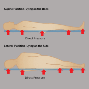
Pressure ulcers develop when sustained pressure restricts blood flow to the skin and tissues. Without adequate blood flow, the affected area is deprived of oxygen and nutrients, leading to tissue damage and the eventual formation of sores. Pressure ulcers commonly occur in areas with bony protrusions, such as the heels, hips, tailbone (sacrum), and elbows. These ulcers can penetrate deep into the muscle and bone in severe cases, causing serious infections.
Pressure Points
Factors Contributing to Pressure Ulcers
Several factors increase the risk of developing pressure ulcers, especially in patients who are bedridden or have limited mobility. These factors include:
1) Immobility
Patients who cannot move independently are at the highest risk. When specific body areas are subjected to prolonged pressure, it reduces circulation and tissue damage.
2) Moisture
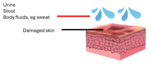
Excess moisture from sweat, urine, or wound drainage can weaken the skin’s protective barrier, making it more susceptible to breakdown.
Moisture-associated skin damage
3) Friction and Shear
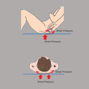
Friction occurs when the skin rubs against a surface, while shear happens when the skin moves in one direction, and the underlying tissue moves in another. Both can contribute to skin damage and ulcer formation.
Direct Pressure and Shear Pressure
4) Poor Nutrition
Malnutrition and dehydration impair the body’s ability to heal and maintain healthy skin, increasing the risk of ulcers.
Underlying Health Conditions: Conditions such as diabetes, vascular disease, and incontinence can exacerbate the risk of pressure ulcers due to poor circulation, reduced sensation, and frequent exposure to moisture.
Key Prevention Strategies
Regular Repositioning
To prevent pressure ulcers, it’s crucial to reposition bedridden patients regularly. Shifting the patient’s position every two hours can relieve pressure on vulnerable areas and promote healthy blood flow. Creating a consistent repositioning schedule is crucial to enhance overall comfort and prevent pressure ulcers.
Always use lifting devices or draw sheets to minimize friction and shear forces when repositioning. Avoid dragging the patient across the bed, which can cause skin damage. Instead, gently lift and reposition them to ensure smooth movement.
Use Supportive Surfaces
Investing in pressure-relieving surfaces, such as specialized mattresses and cushions, can significantly help prevent ulcers. These surfaces are designed to distribute weight evenly and reduce pressure on high-risk areas. Options include foam mattresses, air mattresses, and gel cushions, each offering different levels of support.
Pressure-relieving mattresses can automatically adjust to the patient’s movements, ensuring continuous pressure redistribution. For wheelchair users, pressure-relieving cushions can provide similar benefits by reducing the risk of sores on the buttocks and thighs.
Maintain Skin Hygiene
Proper skin hygiene is crucial in preventing pressure ulcers. The skin should be kept clean and dry to prevent breakdown. Moisture from sweat, urine, or wound drainage can weaken the skin, making it more prone to sores. Use mild, pH-balanced cleansers to wash the skin, and ensure it is thoroughly dried after cleansing.
Applying moisture barriers or creams can protect the skin from excessive moisture, especially in areas prone to incontinence. These products create a protective layer that shields the skin from irritants and helps maintain its integrity.
Ensure Proper Nutrition
Nutrition plays a vital role in skin health and preventing pressure ulcers. A well-balanced diet rich in protein, vitamins, and minerals supports the body’s ability to heal and regenerate tissues. Patients should be encouraged to consume adequate calories and protein to maintain their strength and immune function.
Specific nutrients, such as vitamin C, zinc, and omega-3 fatty acids, are particularly beneficial for skin repair. Hydration is equally important; adequate fluid intake helps maintain skin elasticity and prevents dehydration, which can contribute to skin breakdown.
Regular Skin Inspections
Regular skin inspections are essential for the early detection of pressure ulcers. Caregivers should thoroughly examine the patient’s skin at least once daily, paying close attention to areas where bones are close to the surface. Look for early signs of pressure damage, such as redness, warmth, or changes in skin texture.
Immediately alleviate pressure and provide appropriate care if any suspicious areas are identified. Early intervention can prevent minor skin irritations from developing into full-blown ulcers, significantly reducing the risk of complications.
Managing Incontinence
Incontinence can contribute to pressure ulcer development due to constant skin exposure to moisture and irritants. Managing incontinence effectively is essential for preventing ulcers. Use absorbent pads or briefs that wick moisture away from the skin, and change them frequently to keep the skin dry.
In addition, cleanse the skin gently after each episode of incontinence and apply moisture barriers to protect against irritation. Managing incontinence is an ongoing process that requires attention to detail and consistent care.
Education and Training
Educating patients, caregivers, and healthcare providers about pressure ulcer prevention is crucial. Proper training ensures that everyone involved in the patient’s care knows the risks and how to implement preventive measures. Providing educational materials and regular training sessions can empower caregivers to take proactive steps in preventing pressure ulcers.
Conclusion
Preventing pressure ulcers in bedridden patients is a multifaceted approach that requires diligence, knowledge, and consistent care. Caregivers can significantly reduce the risk of pressure ulcers by incorporating regular repositioning, using supportive surfaces, maintaining skin hygiene, ensuring proper nutrition, and conducting regular skin inspections. Additionally, managing incontinence and providing education and training further enhance prevention efforts. Taking these proactive steps prevents pressure ulcers and improves patients’ overall quality of life.
Learn about cellulitis of the leg: causes, symptoms, treatment options, and prevention tips to manage and avoid this serious skin infection.
Leg Cellulitis: Causes, Symptoms, and Treatments
Introduction
Cellulitis is a common, potentially serious bacterial skin infection. It can cause significant pain, swelling, and discomfort when it affects the leg. This blog will explore the causes, symptoms, and treatments of leg cellulitis, providing valuable information for those affected by this condition.
What is Cellulitis?
Cellulitis is an infection of the deeper layers of the skin, specifically the dermis and subcutaneous tissue. It can occur when bacteria enter the skin through a cut, scrape, or break. Bacteria like Streptococcus and Staphylococcus commonly cause it. If left untreated, cellulitis can rapidly progress and spread to other body parts.
If you suspect cellulitis, it’s essential to seek medical attention. Prompt diagnosis and treatment can prevent the infection from worsening and potentially leading to more severe complications.
Causes of Cellulitis in the Leg
Several factors can lead to the development of cellulitis in the leg:
1) Breaks in the Skin:
Cuts, scrapes, insect bites or surgical wounds can allow bacteria to enter the body.
Skin scratches
Insect bites
2) Chronic Skin Conditions:
Conditions like eczema or athlete’s foot can create openings in the skin.
Eczema
3) Weakened Immune System:
Individuals with compromised immune systems, such as diabetes, are at a higher risk.
There are various conditions that can cause leg swelling, for example, chronic venous insufficiency/ varicose veins and lymphedema. People with leg swelling or oedema, are prone to cellulitis for several reasons:
a) Reduced Blood Circulation:
Swelling in the legs can reduce blood flow, making it easier for bacteria to thrive and multiply.
b) Skin Breakdown:
Persistent swelling can stretch the skin, making it more fragile and prone to minor injuries or cracks. These
skin breaks can provide entry points for bacteria.
c) Stagnant Fluid:
Oedema often leads to the accumulation of fluid in the tissues. This stagnant fluid can be a breeding
ground for bacteria.
d) Weakened Immune Response:
The immune system may not function as effectively in areas with poor circulation and fluid buildup,
reducing the body’s ability to fight off infections.
Chronic venous insufficiency/ Varicose veins
Lymphedema
Symptoms of Cellulitis
Cellulitis of the leg presents with several distinct symptoms:
1) Redness:
The affected skin may also appear shiny, tight, and tender to the touch. In some cases, red streaks may extend from the affected area, indicating the spread of infection along lymphatic channels. As the disease progresses, individuals may experience systemic symptoms such as fever, chills, malaise, and swollen lymph nodes near the site of infection.
2) Swelling:
Swelling and a feeling of tightness in the skin are common.
3) Pain:
The area is usually tender and painful to the touch.
4) Warmth:
The infected skin may feel warm or hot.
5) Fever:
A person may develop a fever and chills in more severe cases.
In severe cases, untreated cellulitis can lead to complications such as abscess formation, tissue necrosis, sepsis, or the spread of infection to deeper structures like muscles and bones.
Cellulitis that affects individuals with compromised immune systems or underlying medical conditions may progress rapidly and result in life-threatening complications.
Timely administration of appropriate antibiotics and close monitoring of the response to treatment are essential in managing cellulitis and preventing complications.
Diagnosing Cellulitis
A healthcare professional can diagnose cellulitis by physical examination and reviewing the patient’s medical history. In some cases, the doctor may recommend additional tests such as blood or wound cultures or imaging studies like ultrasound to confirm the diagnosis, identify the causative bacteria, and assess the severity of the infection. Prompt and accurate diagnosis of cellulitis is essential for initiating appropriate treatment and preventing the spread of infection.
Differential diagnosis: (conditions that mimic cellulitis)
It’s essential to treat cellulitis promptly to prevent the infection from spreading. Treatment options include:
1) Antibiotics:
Treatment options for cellulitis of the leg typically involve using oral or intravenous antibiotics to eradicate the underlying bacterial infection. The severity of the infection, the probable causative bacteria, and any underlying medical conditions that may impact treatment determine the choice of antibiotic therapy. In mild cases of cellulitis, oral antibiotics oral antibiotics may suffice. For more severe infections or cases of antibiotic resistance, intravenous antibiotics administered in a hospital setting may be necessary.
2) Pain Relief:
Over-the-counter pain relievers can help manage discomfort.
Reducing swelling will help to alleviate symptoms and promote healing.
4) Wound Care:
Proper cleaning and care of open wounds are essential to prevent further infection and aid in the resolution of cellulitis.
Wound care
Preventing Cellulitis
Preventing cellulitis involves taking measures to protect the skin from injury and infection:
1)Proper wound care:
This includes promptly cleaning and covering any cuts or wounds, which can prevent bacteria from entering the skin and causing infection.
2) Good Hygiene:
Maintaining good hygiene practices, such as washing and moisturizing hands regularly, showering after physical activities, and keeping living spaces clean, can help prevent the spread of bacteria that cause skin infections.
3) Protective Clothing:
Wearing protective gear during activities that risk skin injury.
4) Prompt Treatment:
Promptly treat cuts, scrapes, and other skin injuries to prevent infection.
Conclusion
Cellulitis of the leg is a common skin infection that can cause significant discomfort and complications if left untreated. Understanding the causes, symptoms, and treatment options for cellulitis is essential for prompt recognition and appropriate management. Prompt diagnosis and treatment of cellulitis are crucial for preventing complications and ensuring optimal outcomes.
It is essential to differentiate cellulitis from other skin conditions like eczema, contact dermatitis, or venous stasis dermatitis, as the treatment approach may vary. Seeking medical evaluation for persistent or worsening skin changes is essential for accurate diagnosis and appropriate management of cellulitis.
Discover the causes, symptoms, diagnosis, and treatments for Pelvic Congestion Syndrome, a commonly misunderstood cause of chronic pelvic pain in women.
Introduction
Pelvic congestion syndrome (PCS) is a common yet often misunderstood cause of chronic pelvic pain in women. The presence of varicose veins in the pelvis, which causes pelvic vein insufficiency and a variety of distressing symptoms, is what distinguishes this condition. In this blog, we will explore the causes, symptoms, diagnosis, and treatment options for pelvic congestion syndrome.
Pelvic congestion syndrome occurs when veins in the pelvic region become enlarged and engorged, leading to chronic pelvic pain. Similar to varicose veins in the legs, these pelvic varicose veins fail to adequately return blood to the heart, resulting in blood pooling in the veins and causing discomfort.
Symptoms of Pelvic Congestion Syndrome
The symptoms of PCS can include a range of symptoms, including:
1) Chronic pelvic pain
The hallmark symptom of PCS is chronic pelvic pain that lasts six months or longer. This pain is often described as a dull ache in the lower abdomen, but it can also be sharp, throbbing, or cramp-like. It may worsen with prolonged sitting or standing and be relieved by lying down.
2) Lower back pain
Pressure from engorged varicose veins due to blood pooling and the chronic inflammation associated with PCS can extend to surrounding areas, including the lower back. Inflammation can irritate the nerves and tissues, causing pain and discomfort in the lower back region.
3) Varicose veins in the pelvic area
These are enlarged and twisted veins that may be visible through the skin, especially in the vulva or buttocks area.
4) Pain during or after sexual intercourse
Some individuals with PCS may experience pain during or after sexual intercourse. This can be due to increased blood flow to the pelvic region during arousal, which exacerbates the symptoms of PCS.
5) Irritable bladder and bowel symptoms
Irritation of bladder and bowel from swollen pelvic veins can lead to:
bladder spasms and a strong urge to urinate
Frequent bouts of diarrhoea alternating with constipation
6) Worsening of symptoms during menstruation
For some individuals, PCS symptoms may worsen during their menstrual cycle. This is due to hormonal changes that increase the blood flow and volume in the pelvic region.
Causes of Pelvic Congestion Syndrome
The exact cause of pelvic congestion syndrome is not entirely understood, but several factors are believed to contribute to the condition:
1) Hormonal changes:
High oestrogen levels, particularly during pregnancy, can weaken vein walls, leading to varicose veins.
2) Multiple pregnancies:
Increased blood flow and pressure during pregnancy can cause pelvic veins to enlarge.
3) Genetics:
may also play a role in the development of PCS. Some individuals may be predisposed to weak vein valves, absence of valves or other abnormalities in their blood vessels, making them more susceptible to developing this condition.
4) Secondary pelvic vein incompetence
This is a condition where the veins in the pelvis are unable to efficiently transport blood back to the heart due to external compression. The nutcracker syndrome, also known as left renal vein entrapment syndrome, is one of many factors that can cause this external compression. This phenomenon occurs when the left renal vein, which carries blood from the left kidney to the heart, is compressed between the superior mesenteric artery and the abdominal aorta. Similarly, May-Thurner syndrome can also lead to pelvic vein incompetence due to the compression of the left common iliac vein by the right internal iliac artery.
Diagnosing Pelvic Congestion Syndrome
Diagnosing PCS can be challenging due to the overlap of symptoms with other conditions. A thorough medical history and physical examination are essential. Diagnostic tests that may be used include:
A) Pelvic ultrasound:
Pelvic ultrasound is the first-line imaging study for PCS. This non-invasive imaging allows healthcare providers to visualize any abnormalities in the pelvic veins. It can also rule out the presence of pelvic masses or uterine problems as the underlying cause of pelvic pain
Pelvic ultrasound scan: dilated pelvic veins
B) Computed Tomography Venogram (CTV) and Magnetic Resonance Venogram (MRV)
CTV and MRV are useful for providing detailed images of pelvic structures and blood vessels. They can rule out pelvic masses and compression of the veins in the abdomen and pelvis. They are both minimally invasive imaging techniques with low risk.
MRI of the abdomen and pelvis: dilated left ovarian vein and pelvic varicose vein
CT scan: varicose veins in the pelvis
C) Contrast Venography:
Contrast venography is the gold standard for diagnosing pelvic congestion. This procedure involves injecting contrast dye into the pelvic veins and taking X-ray images to identify any areas of blood pooling or vein abnormalities. Venograms provide a more detailed view of the pelvic veins and can help guide treatment decisions.
The treatment options for pelvic congestion syndrome can vary depending on the severity of symptoms and individual patient factors. The goal of treatment is to alleviate pain, improve quality of life, and reduce the risk of complications associated with PCS.
1. Conservative management:
In mild cases of PCS, conservative management may be recommended as a first-line treatment. This approach includes lifestyle modifications such as regular exercise, avoiding prolonged sitting or standing, and wearing compression stockings to improve blood flow in the legs and pelvis.
2. Medications:
Painkillers such as nonsteroidal anti-inflammatory drugs (NSAIDs) may be recommended to help manage pelvic pain associated with PCS. Hormonal medications, such as birth control pills or hormonal therapy, may also be prescribed to regulate hormone levels and reduce symptoms.
3. Minimally invasive procedures:
For individuals with more severe symptoms, minimally invasive procedures, namely Ovarian Vein Embolization, may be recommended to alleviate pain and improve blood flow in the pelvic veins.
In this procedure, a catheter (tube) is placed directly into the abnormal ovarian and pelvic veins. Sclerosing agents (chemicals that induce irritation or inflammation) are injected into the pelvic varicose veinsand cause them to collapse. Small metal coils or plugs are then placed to block flow into the ovarian vein. This prevents the reversal of flow in the abnormal veins, which reduces the pressure within the enlarged pelvic veins.
Embolization of left ovarian vein
4. Surgical intervention:
In rare cases, surgical intervention may be necessary to remove or repair damaged veins or valves in the pelvic region. This option is typically reserved for individuals with severe and refractory symptoms who have not responded to other treatments.
It is important to consult with a healthcare provider who specialises in the management of pelvic pain and vascular conditions to determine the most appropriate treatment approach for your situation.
Conclusion
Pelvic congestion syndrome is an often overlooked and misunderstood condition that causes chronic pelvic pain. By understanding the causes, symptoms, and available treatment options for PCS, individuals can take proactive steps towards finding relief and improving their quality of life.
To address chronic pelvic pain or suspect pelvic congestion syndrome, consult with a healthcare provider specialising in pelvic pain or vascular conditions. They can assist in diagnosing the condition and directing you towards a suitable treatment option
Remember, you are not alone in your journey with pelvic congestion syndrome. There is support available, and with the right treatment and lifestyle modifications, you can manage your symptoms and reclaim your life from the grip of this invisible illness. Don’t let pelvic congestion syndrome silence your voice. Seek the help you deserve and take the first step towards finding relief today.